

 PDF
PDF ePub
ePub Citation
 Print
Print


Abstract
As radiation therapy is one of three major cancer treatment methods, many cancer patients get radiation therapy. To exposure as much radiation to cancer while normal tissues near tumor get little radiation, medical physicists make a radiotherapy plan treatment and perform quality assurance before patient treatment. Despite these efforts, unintended medical accidents can occur by some errors. In order to solve the problem, patient internal dose reconstruction methods by measuring transit dose are suggested. As feasibility study for development of patient dose verification system, inverse square law, percentage depth dose and scatter factor are used to calculate dose in the water-equivalent homogeneous phantom. As a calibration results of ionization chamber and glass dosimeter to transit radiation, signals of glass dosimeter are 0.824 times at 6 MV and 0.736 times at 10 MV compared to dose measured by ionization chamber. Average scatter factor is 1.4 and Mayneord F factor was used to apply percentage depth dose data. When we verified the algorithm using the water-equivalent homogeneous phantom, maximum error was 1.65%.
Go to : 
References
1. Hall EJ, Wuu CS. Radiation-induced second cancers: the impact of 3D-CRT and IMRT. International Journal of Radiation Oncology Biology Physics. 56(1):83–88. 2003.

2. Kim S, Min BJ, Yoon M, et al. Secondary radiation doses of intensitymodulated radiotherapy and proton beam therapy in patients with lung and liver cancer. Radiotherapy and oncology: Journal of the European Society for Therapeutic Radiology and Oncology. 98(3):335–339. 2011.
3. Kim D, Sung J, Lee H, et al. Estimation of Secondary Scattered Dose from Intensity-modulated Radiotherapy for Liver Cancer Cases, Progress in Medical Physics. 24(4):295–302.
4. Boellaard R, Essers M, Van Herk M, Mijnheer BJ. New method to obtain the midplane dose using portal in vivo dosimetry. International Journal of Radiation Oncology Biology Physics. 41(2):465–474. 1998.
5. Kasper LP, Marco K, Sandra Q, Andries GV, Ben JM Heijmen: Transit dosimetry with an electronic portal imaging device(EPID) for 115 prostate cancer patients. International Journal of Radiation Oncology Biology Physics. 45(5):1297–1303. 1999.
6. Rascal F, Philippe B, Lucie B, Alejandro M. In vivo dose verification from back projection of a transit dose measurement on the central axis of photon beams. Physica Medica. 27(1):1–10. 2011.
7. S Nijsten, W Elmpt, M Jacobs, et al. A global calibration model for a-Si EPIDs used for transit dosimetry. Medical Physics. 34(10):3872–3884. 2007.
8. R Bogaerts, AV Esch, Rita Reymen, D Huyskens: A method to estimate the transit dose on the beam axis for verification of dose delivery with portal images. Radiotherapy and Oncology. 54(1):39–46. 2000.
9. Cilla S, Meluccio D, Fidanzio A, et al. Initial clinical experience with Epid-based in-vivo dosimetry for VMAT treatments of head-and-neck tumors. Physica Medica (. 2015.
10. BJ Mijnheer, P Gonzalez, IO Ruiz, et al. Overview of 3-year experience with large-scale electronic portal imaging device-based 3-dimensional transit dosimetry. Practical Radiation Oncology. 5(6):679–687. 2015.
11. LCGG Persoon, M Podesta, L Hoffmann, et al. Is integrated transit planar portal dosimetry able to detect geometric changes in lung cancer patients treated with volumetric modulated arc therapy? Acta Oncologica. 54(9):1501–1507. 2015.
12. Khan FM. The Physics of Radiation Therapy. 3rd ed., Williams & Wilkins, Baltimore, MD (. 2003. ), pp.158–177.
13. IAEA TRS-398: Absorbed Dose Determination in External Beam Radiotherapy: An International Code of Practice for Dosimetry based on Standards of Absorbed Dose to Water. International Atomic Energy Agency (. 2000.
Go to :
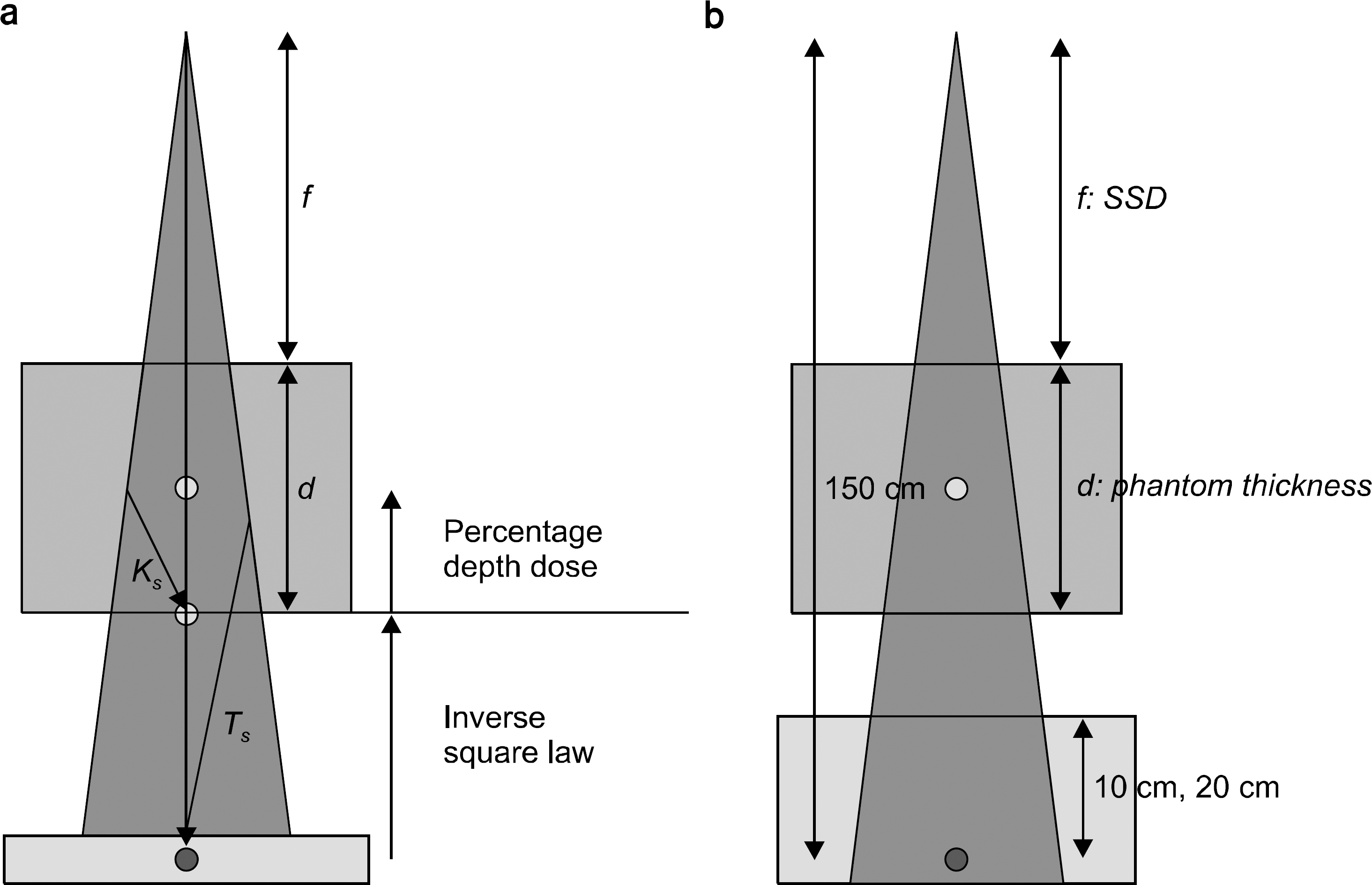 | Fig. 1.(a) The outline of experimental setting for dose calculation in the homogeneous phantom using the transit dose and (b) beam quality correction method of ionization chamber using IAEA TRS-398. Each Ks, Ts, f and d represent an amount of scattered dose to the bottom point, an amount of scattered dose to the transit dose measurement point, source to surface distance (SSD) and phantom thickness. Phantom dose can be calculated by measured transit dose multiplied by inverse square law factor and percentage depth dose data. |
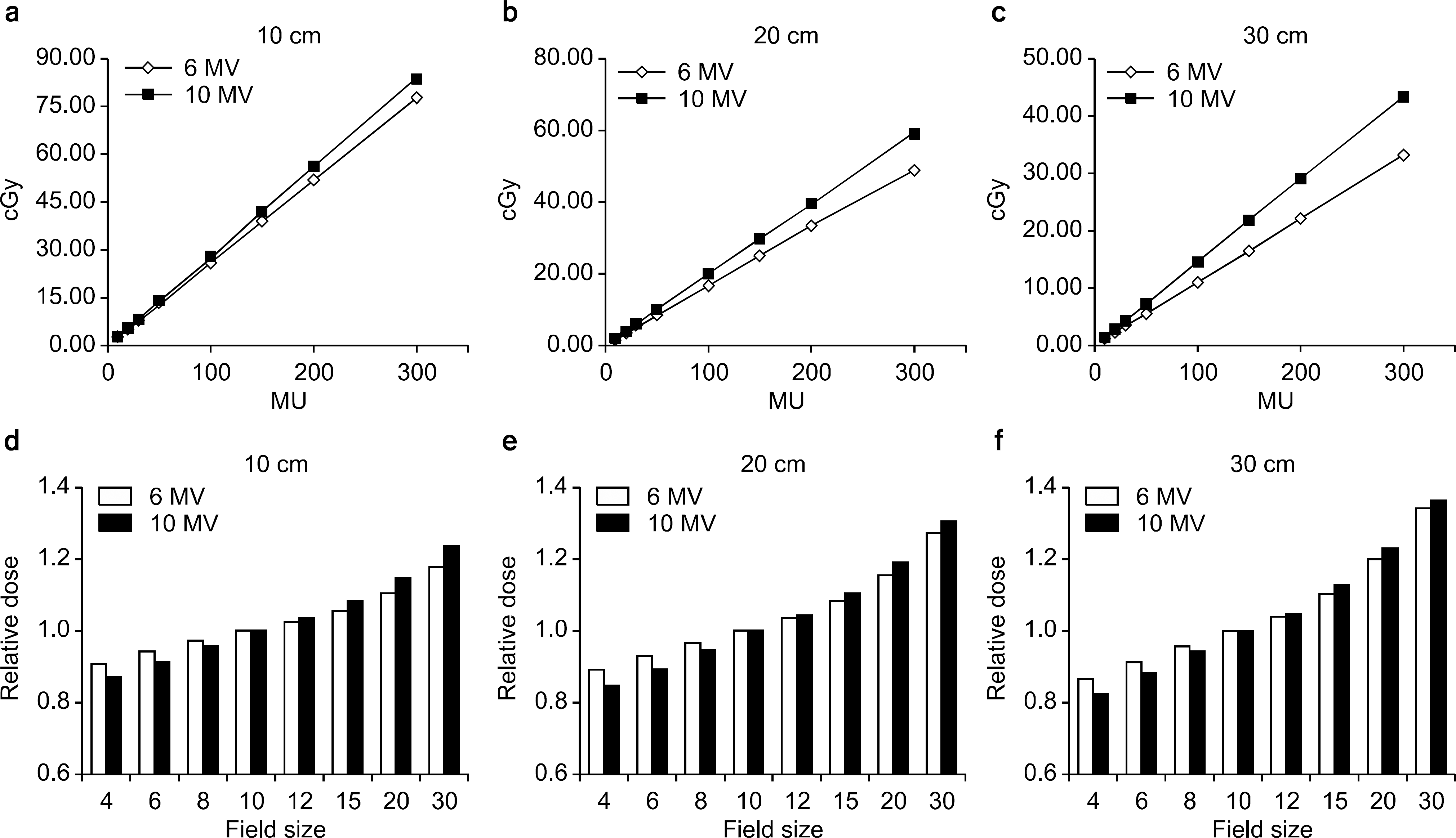 | Fig. 2.Measurement of transit radiation dose according to variation of radiation energy, field size and monitor unit using the correction completed ionization chamber to transit radiation. (a) MU dependency at 10 cm phantom (b) MU dependency at 20 cm phantom (c) MU dependency at 30 cm phantom (d) Field size dependency at 10 cm (e) Field size dependency at 20 cm (f) Field size dependency at 30 cm. |
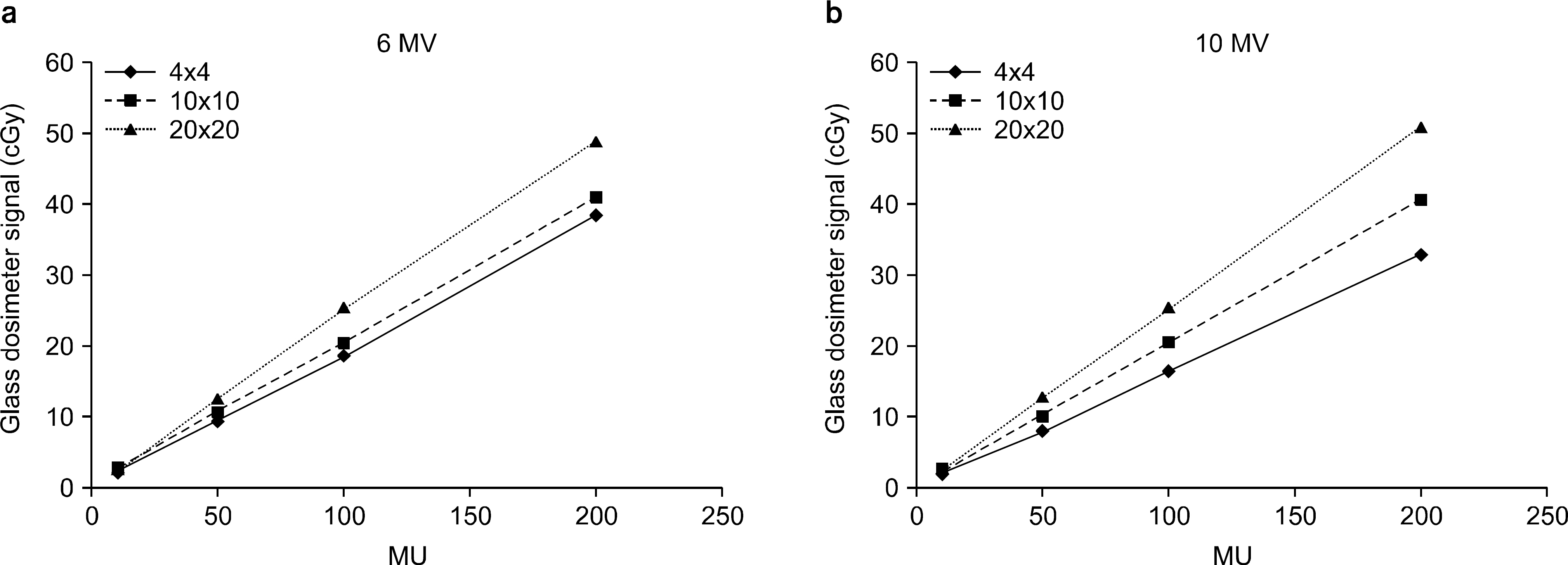 | Fig. 3.(a) 6 MV transit dose measurement results and (b) 10 MV transit dose measurement results measured by glass dosimeter of field size 4 cm×4 cm (◆), 10 cm×10 cm (■), 20 cm×20 cm (▲). |
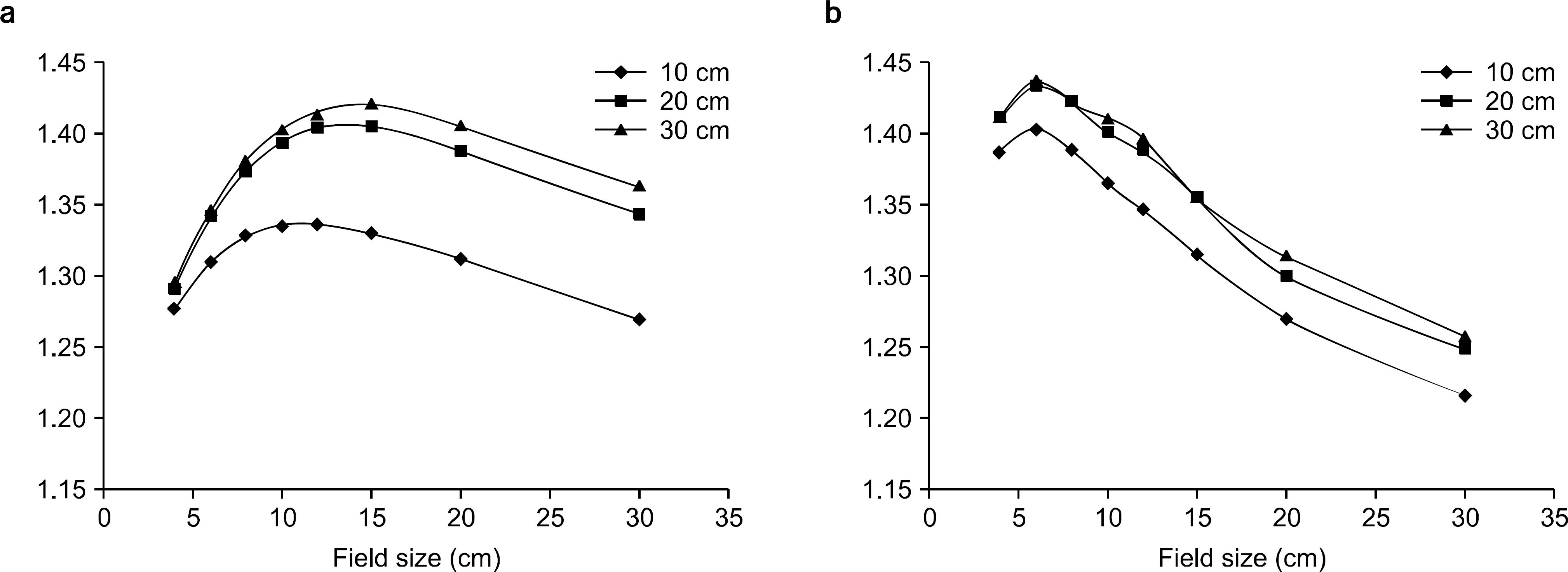 | Fig. 4.Scatter factor measurement results of (a) 6 MV photon energy and (b) 10 MV photon energy when phantom thicknesses are 10 cm (◆), 20 cm (■), 30 cm (▲). |
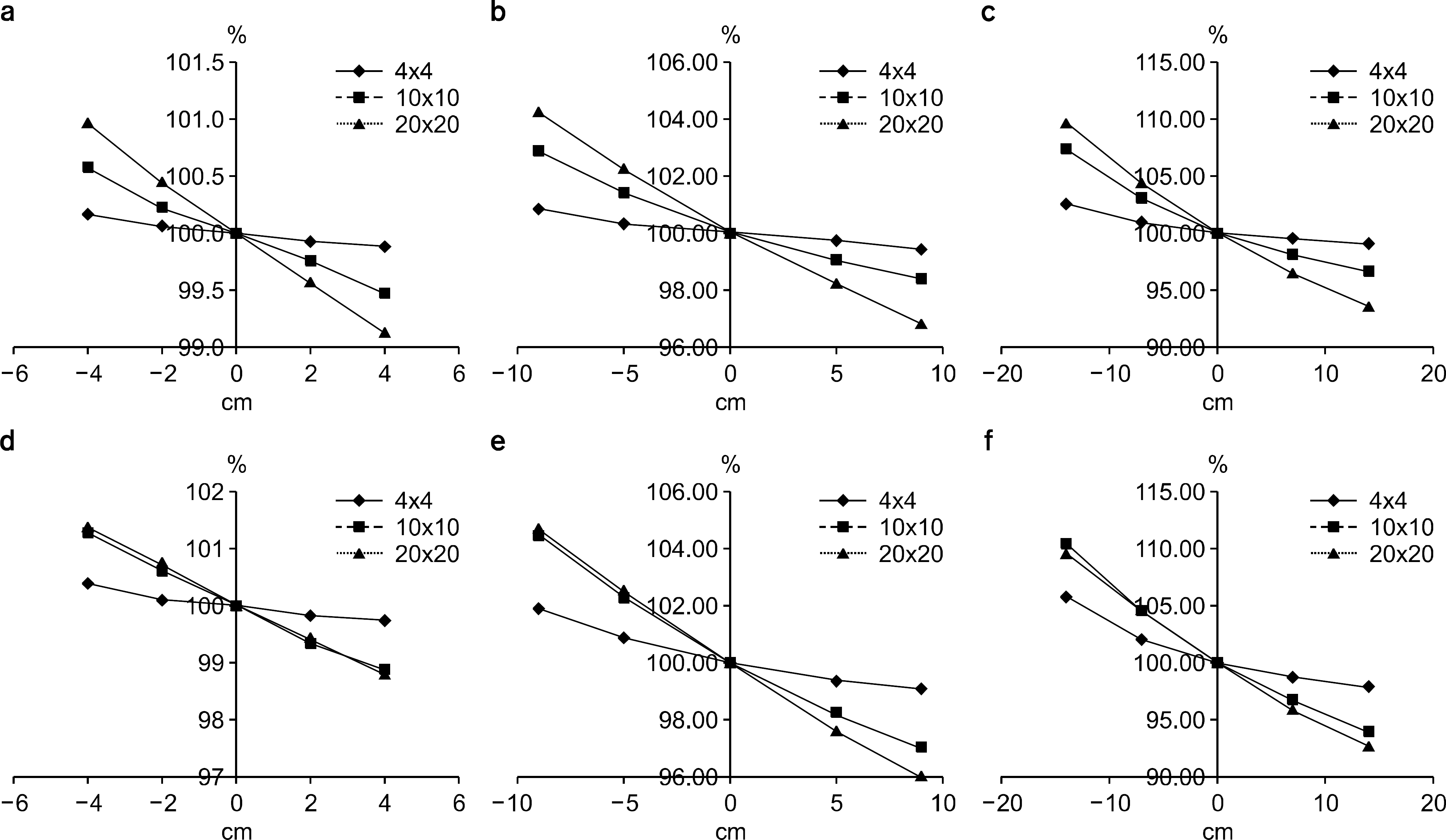 | Fig. 5.Dose variation due to positional change of phantom center to isocenter. Relative dose change when the phantom center is above the isocenter (positive side of horizontal axis), below the isocenter (negative side of horizaontal axis) and on the isocenter (0). The results of (a) 6 MV 10 cm, (b) 6 MV 20 cm, (c) 6 MV 30 cm, (d) 10 MV 10 cm, (e) 10 MV 20 cm, (f) 10 MV 30 cm when field sizes are 4 cm×4 cm (◆), 10 cm×10 cm (■), 20 cm×20 cm (▲). |
Table 1.
The comparison of ionization chamber correction between transit dose result and original TRS-398 result.
Table 2.
The comparison between real measurement results at the center and bottom of 10 cm, 20 cm, 30 cm phantom and calculation results using the Mayneord F factor.
Table 3.
Measurement result of transit radiation and comparison of calculation results using the 6 MV photon.
 XML Download