Abstract
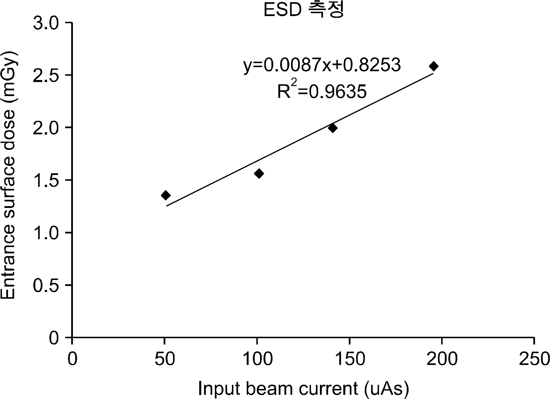
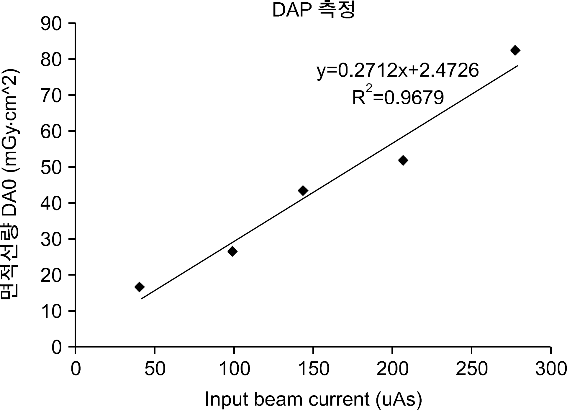
Radiation exposure from medical diagnostic imaging procedures to patients is one of the most significant interests in diagnostic x-ray system. A miniature x-ray intraoral tube was developed for the first time in the world which can be inserted into the mouth for imaging. Dose evaluation should be carried out in order to utilize such an imaging device for clinical use. In this study, dose evaluation of the new x-ray unit was performed by 1) using a custom made in vivo Pig phantom, 2) determining exposure condition for the clinical use, and 3) measuring patient dose of the new system. On the basis of DRLs (Diagnostic Reference Level) recommended by KDFA (Korea Food & Drug Administration), the ESD (Entrance Skin Dose) and DAP (Dose Area Product) measurements for the new x-ray imaging device were designed and measured. The maximum voltage and current of the x-ray tubes used in this study were 55 kVp, and 300 mA. The active area of the detector was 72×72 mm with pixel size of 48 μm. To obtain the operating condition of the new system, pig jaw phantom images showing major tooth-associated tissues, such as clown, pulp cavity were acquired at 1 frame/sec. Changing the beam currents 20 to 80 μA, x-ray images of 50 frames were obtained for one beam current with optimum x-ray exposure setting. Pig jaw phantom images were acquired from two commercial x-ray imaging units and compared to the new x-ray device: CS 2100, Carestream Dental LLC and EXARO, HIOSSEN, Inc. Their exposure conditions were 60 kV, 7 mA, and 60 kV, 2 mA, respectively. Comparing the new x-ray device and conventional x-ray imaging units, images of the new x-ray device around teeth and their neighboring tissues turn out to be better in spite of its small x-ray field size. ESD of the new x-ray device was measured 1.369 mGy on the beam condition for the best image quality, 0.051 mAs, which is much less than DRLs recommended by IAEA (International Atomic Energy Agency) and KDFA, both. Its dose distribution in the x-ray field size was observed to be uniform with standard deviation of 5∼10 %. DAP of the new x-ray device was 82.4 mGy∗cm2 less than DRL established by KDFA even though its x-ray field size was small. This study shows that the new x-ray imaging device offers better in image quality and lower radiation dose compared to the conventional intraoral units. In additions, methods and know-how for studies in x-ray features could be accumulated from this work.
Go to : 
References
2. Altug HA, Ozkan A. Diagnostic imaging in Oral and Maxillofacial Pathology. Medical Imaging, Eribdy OF, InTech (. 2011. ), pp.215–238.
3. Mihailova Hr, Nikolov Vl, Slavkov Sv: Diagnostic imaging of dentigerous cysts of the mandible. Journal of IMAB. 2:8–10. 2008.
4. Tyndall DA, Rathore S. Cone-Beam CT diagnostic applications: caries, periodontal bone assessment, and endodontic applications. Dent Clin North Am. 52:825–841. 2008.

5. AAPM Report No. 31: Standardized methods for measuring diagnostic x-ray exposures. the American Association of Physicists in Medicine, New York (. 1990.
6. Rivard MJ, Davis SD, DeWerd LA, et al. Calculated and measured brachytherapy dosimetry parameters in water for the Xoft Axxent X-ray Source: an electronic brachytherapy source. Med Phys. 33:4020–4032. 2006.
7. Cho S, Kim D, Baek K, et al. Introduction of dental x-ray imaging with new concept – intra Oral x-ray Tube. J Inst Electron Eng Korea 48, No. 4:94–101. 2011.
8. Cho S, Rena L. The Characteristic of Temperature and Dose Distribution of intra oral X-ray Tube. J Inst Electron Eng Korea. 50(5):62–266. 2013.

9. Cho BC, Huh HD, Kim JS, et al. Guideline for Imaging Dose on Image-Guided Radiation Therapy. Pro. Med Phys. 24(1):1–24. 2013.

10. ICRP: Diagnostic reference levels in medical imaging: Review and additional advice. 2001 Annual Report of the international Commission on Radiological Protection, Hague, (. 2001.
11. IAEA: International Basic Safety Standards for Protection against Ionizing Radiation and for the Safety of Radiation Sources. IAEA safety series No. 115, Vienna, (. 1996.
12. ICRP: Radiological Protection and Safety in Medicine. ICRP Publication 73, Oxford (. 1996.
13. Kim EK. Development of diagnostic reference level in dental x-ray examination in Korea. Final report. Korea Food & Drug Administration, Seoul (. 2009.
Go to : 
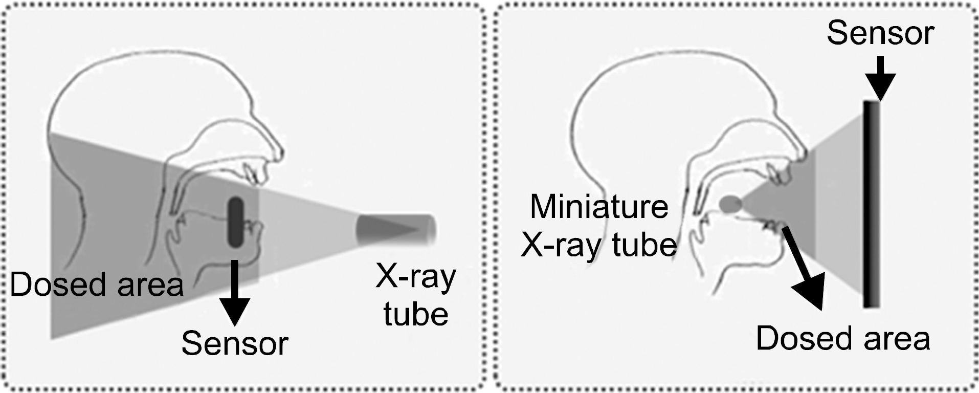 | Fig. 2.Comparison of two x-ray acquisition methods: typical settings for conventional intraoral x-ray unit (Left). Totally different settings for the new conceptual intraoral x-ray unit (Right). |
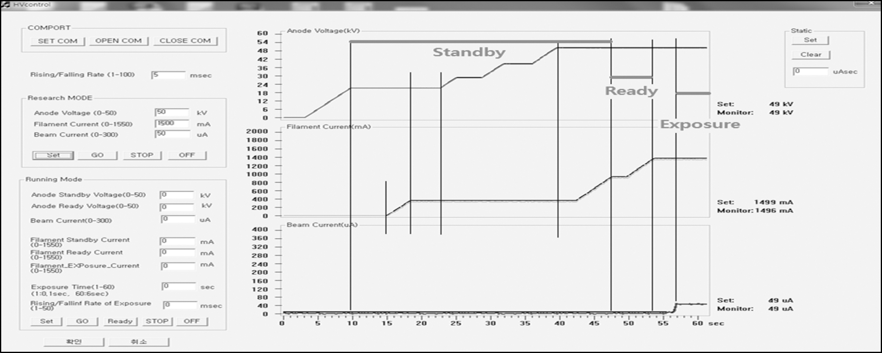 | Fig. 3.The custom-made program developed for the control of tube voltage, beam current, and filament current. |
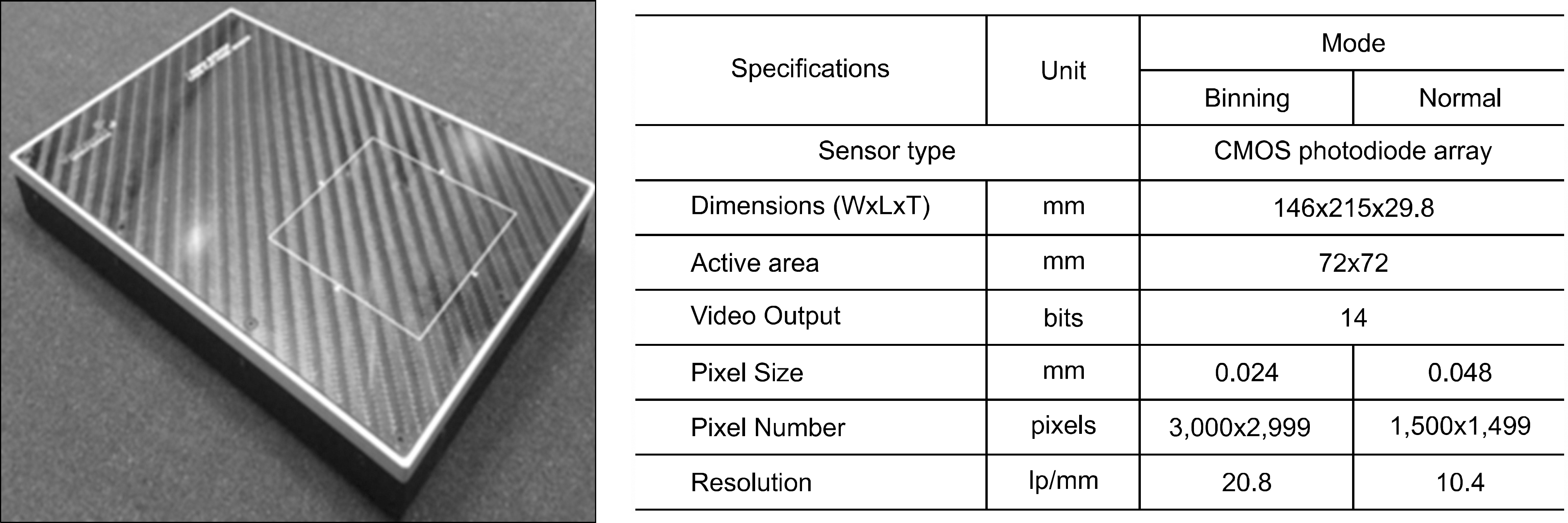 | Fig. 4.Specification of the detector used in this study for the intensity measurements and image acquisition. |
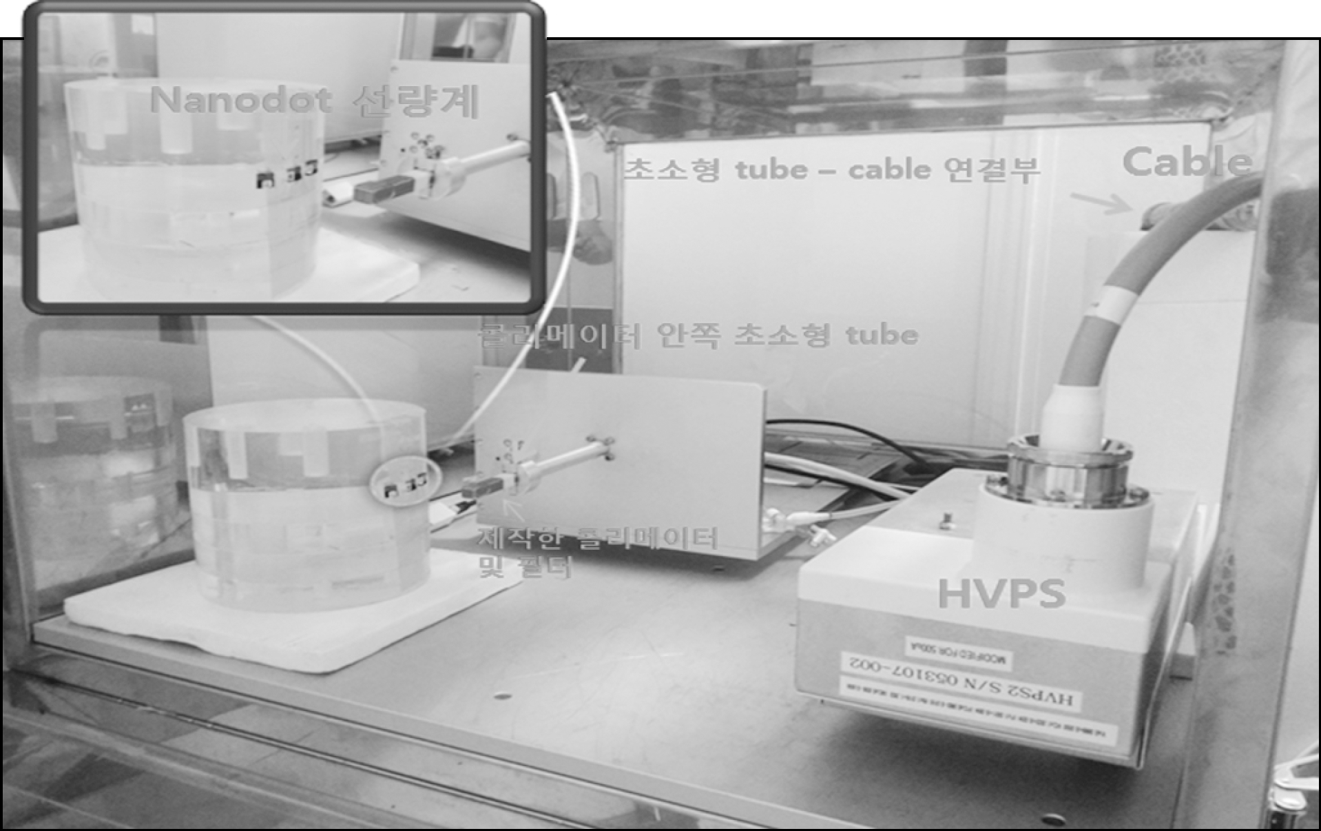 | Fig. 5.The set-up for ESD measurements in an x-ray shielding jig: OSLDs are placed on the surface of the cylindrical phantom. The miniature x-ray tube is located 5 cm away from the OSLDs. |
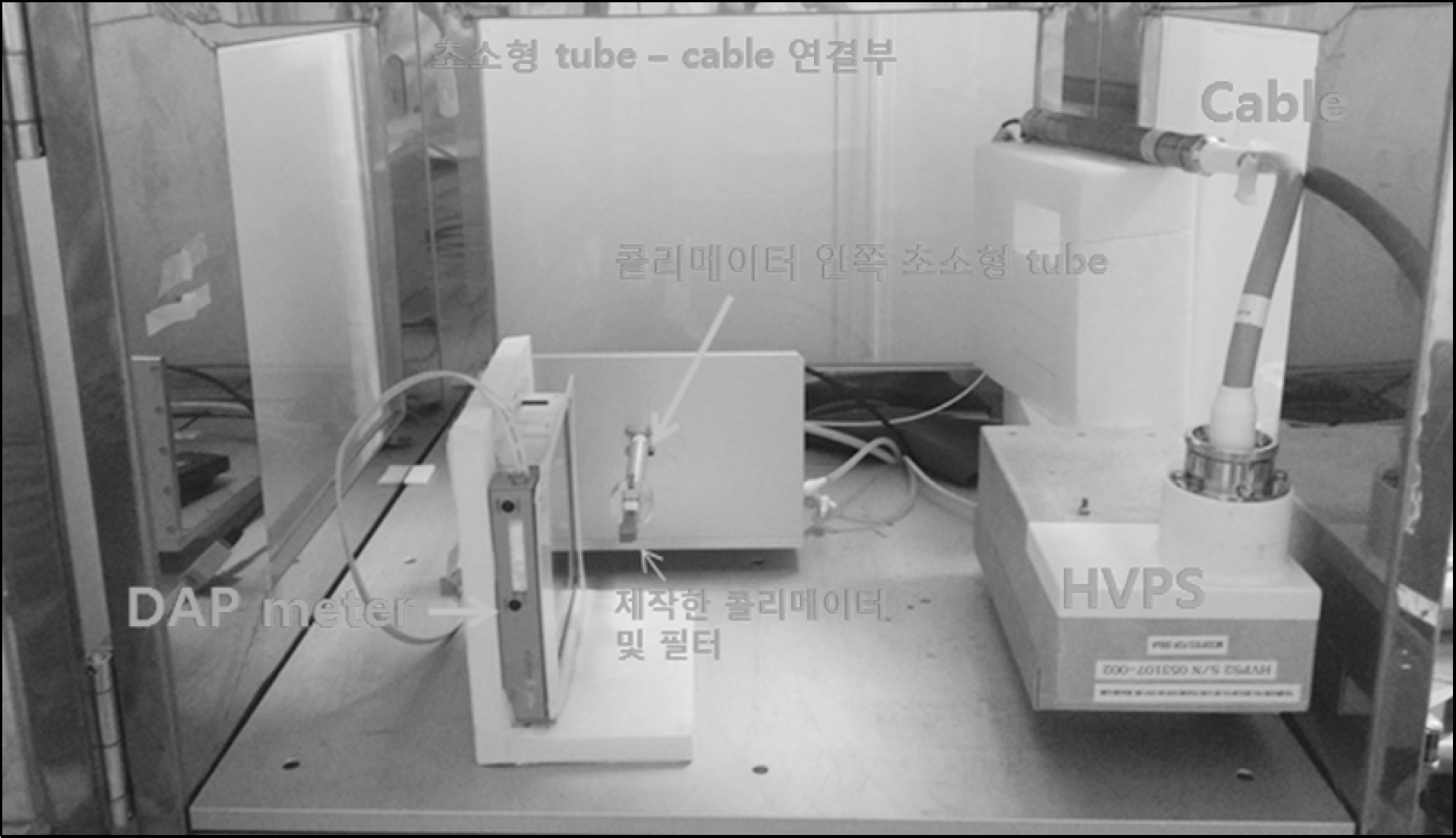 | Fig. 6.The set-up for DAP measurements in an x-ray shielding jig: a DAP meter is placed from the miniature x-ray tube. |
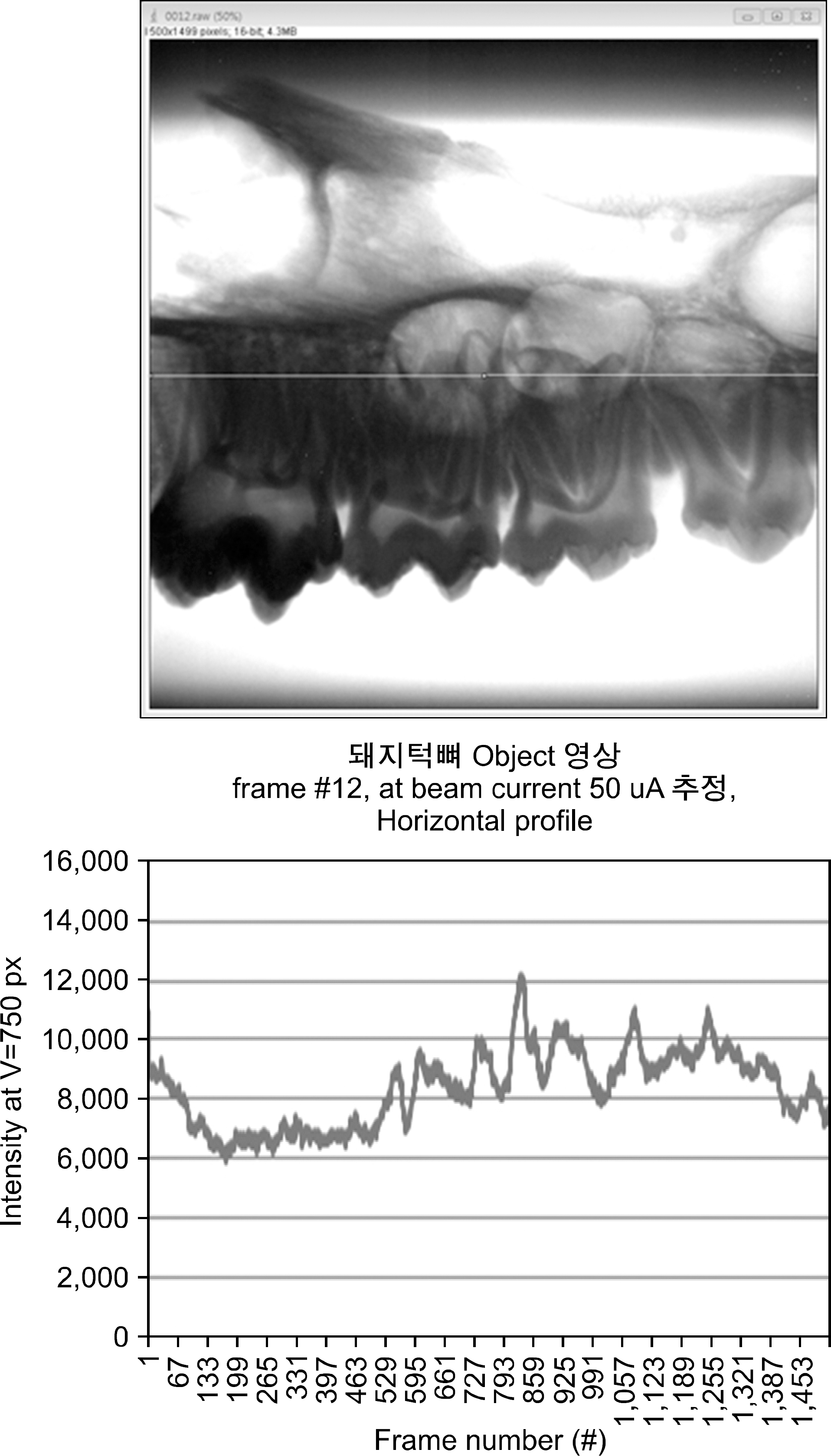 | Fig. 8.X-ray image of pig mandibular molars with intensitiy profile across the image at 50 kVp, 0.051 mAs. |
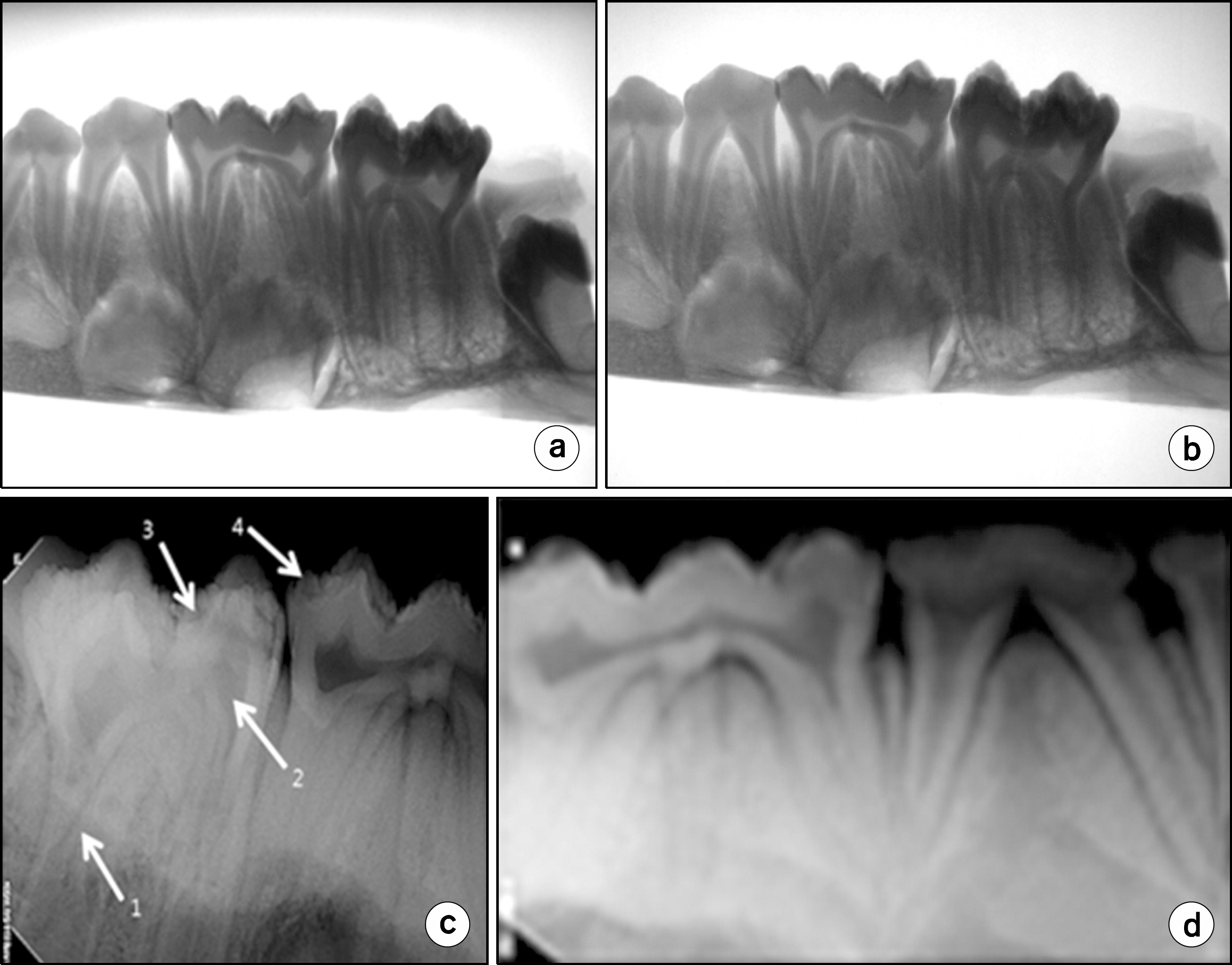 | Fig. 9.X-ray images of the pig mandibular phantom obtained from (a) new system at 50 kV, 40 uA, (b) new system at 55 kV, 30uA, (c) commercial system at 60 kV, 7 mA (CS2100, &RVG digital sensor, Carestream Dental, LLC.) and (d) commercial system at 60 kV, 2 mA (EXARO&EZ digital sensor, Osstem, Inc.). |
Table 1.
ESD (Entrance Surface Dose) values measured with OSLD (Optically Stimulated Luminescence Dosimeter) nanoDot on the phantom as a function of beam current.
| ESD | |||
|---|---|---|---|
| OSLD nanodot chip ID | Position | Beam Current (μAs) | ESD (mGy) |
| DN09450725Q | center | 51 | 1.369 |
| DN09400110N | center | 101 | 1.575 |
| DN09410565Y | center | 141 | 2.02 |
| DN10103146P | center | 196 | 2.601 |


 PDF
PDF ePub
ePub Citation
 Print
Print


 XML Download