

 PDF
PDF ePub
ePub Citation
 Print
Print


Abstract
The purpose of this study is to analyze motion-induced dose error generated by each tumor motion parameters of irregular tumor motion in helical tomotherapy. To understand the effect of the irregular tumor motion, a simple analytical model was simulated. Moving cases that has tumor motion were divided into a slightly irregular tumor motion case, a large irregular tumor motion case and a patient case. The slightly irregular tumor motion case was simulated with a variability of 10% in the tumor motion parameters of amplitude (amplitude case), period (period case), and baseline (baseline case), while the large irregular tumor motion case was simulated with a variability of 40%. In the phase case, the initial phase of the tumor motion was divided into end inhale, mid exhale, end exhale, and mid inhale; the simulated dose profiles for each case were compared. The patient case was also investigated to verify the motion-induced dose error in ‘clinical-like' conditions. According to the simulation process, the dose profile was calculated. The moving case was compared with the static case that has no tumor motion. In the amplitude, period, baseline cases, the results show that the motion-induced dose error in the large irregular tumor motion case was larger than that in the slightly irregular tumor motion case or regular tumor motion case. Because the offset effect was inversely proportion to irregularity of tumor motion, offset effect was smaller in the large irregular tumor motion case than the slightly irregular tumor motion case or regular tumor motion case. In the phase case, the larger dose discrepancy was observed in the irregular tumor motion case than regular tumor motion case. A larger motion-induced dose error was also observed in the patient case than in the regular tumor motion case. This study analyzed motion-induced dose error as a function of each tumor motion parameters of irregular tumor motion during helical tomotherapy. The analysis showed that variability control of irregular tumor motion is important. We believe that the variability of irregular tumor motion can be reduced by using abdominal compression and respiratory training.
Go to : 
References
1. Cedric X Yu, David A Jaffray, John W Wong. Theeffects ofintra-fractionorganmotiononthedeliveryofdynamicin-tensitymodulation. Phys. Med. Biol. 43(1):91–104. 1998.
2. Bortfeld , Thomas , Steve B. Jiang, Eike Rietzel, Effects ofmotiononthetotaldosedistribution.Semin.Radiat.Oncol. 14(1):41–51. 2004.
3. Sung Kyu Kim, Min Kyu Kang, Ji Woon Yea, Se An Oh. Dosimetricevaluationofamovingtumortargetinin-tensity-modulatedradiationtherapy(IMRT)forlungcancer patients. J. KoreanPhys. Soc. 63(1):67–70. 2013.
4. B. Kim, J. Chen, T. Kron and. J. Battista: Motion-induced doseartifactsinhelicaltomotherapy.Phys.Med.Biol. 54(19):5707–5734. 2009.
5. M. Klein, S. Gaede and S. Yartsev: Astudyoflongitudinal tumormotioninhelicaltomotherapyusingacylindricalphantom. J.Appl.Clin.Med.Phys. 14(2):52–61. 2013.
6. J. H. Lewis and S. B. Jiang: Atheoreticalmodelforrespi-ratorymotionartifactsinfree-breathingCTscans.Phys.Med. Biol. 54(3):745–755. 2009.
7. M. W. Kissick, J. Fenwick, J. A. James, et al: Thehelical tomotherapythreadeffect.Med.Phys. 32(5):1414–1423. 2005.
8. J. N. Yang, T. R. Mackie, P. Reckwerdt, J. O. Deasy and B. R. Thomadsen: Aninvestigationoftomotherapy beamdelivery.Med.Phys. 24(3):425–436. 1997.
9. Brian Kanagaki, Paul W Read, Janelle A Molloy, James M Larner, Ke Sheng. Amotionphantomstudyonhelicalto-motherapy: thedosimetricimpactsofdeliverytechniqueand motion. Phys. Med. Biol. 52(1):243–255. 2007.
10. A. Schweikard, H. Shiomi and. J. Adler: Respirationtrackinginradiosurgery.Med.Phys. 31(10):2738–2741. 2004.
11. P. Giraud, E. Morvan, L. Claude, et al: RespiratoryGating TechniquesforOptimizationofLungCancer Radiotherapy.J. Thorac.Oncol. 6(12):2058–2068. 2011.
12. Martin J Murphy: Trackingmovingorgansinrealtime.Semin. Radiat.Oncol. 14(1):91–100. 2004.
13. AAPM TG-69 Report. Radiographicfilmformegavoltage beamdosimetry, Sujatha Pai (. 2007.
14. Martina Fuss, Eva Sturtewagen, Carlos De Wagter, Dietmar Georg: DosimetriccharacterizationofGafChromic EBTfilmanditsimplicationonfilmdosimetryqualityassurance. Phys.Med.Biol. 52(14):4211–4225. 2007.
15. Bart D. Lynch, Jakub Kozelka, Manisha K. Ranade, et al: Importantconsiderationsforradiochromicfilmdosimetrywith flatbedCCDscannersandEBTGAFCHROMIC®film. Jonathan G, Li, WilliamE. Simon, James F. Dempsey, Med. Phys. 33(12):4551–4556. 2006.
16. Sarah Quirk, Nathan Becker, and Wendy Smith. External respiratorymotion: Shapeanalysisandcustomrealisticrespiratorytracegeneration. Med. Phys. 39(8):4999–5003. 2012.
17. AAPM TG-76 Report: Themanagement of respiratorymotioninradiationoncology. P.J. Keall. 2006.
Go to :
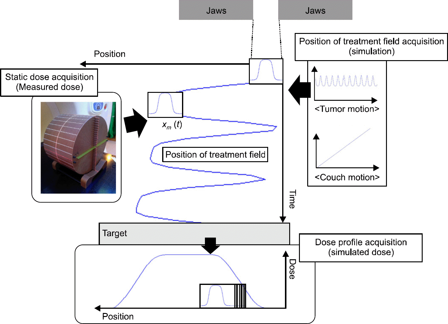
 | Fig. 2.Position versus time curve of the treatment field in the (a) room coordinate system and (b) tumor coordinate system. |
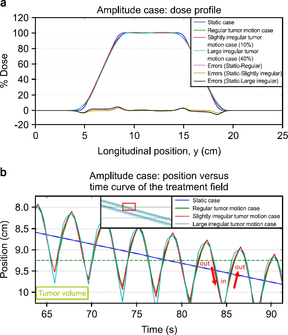 | Fig. 3.(a) Dose profiles in the amplitude case. (b) Position versus time curve in the amplitude case. |
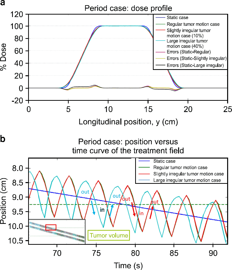 | Fig. 4.(a) Dose profiles in the period case. (b) Position versus time curve in the period case. |
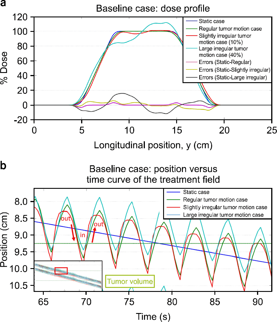 | Fig. 5.(a) Dose profiles in the baseline case. (b) Position versus time curve in the baseline case. |
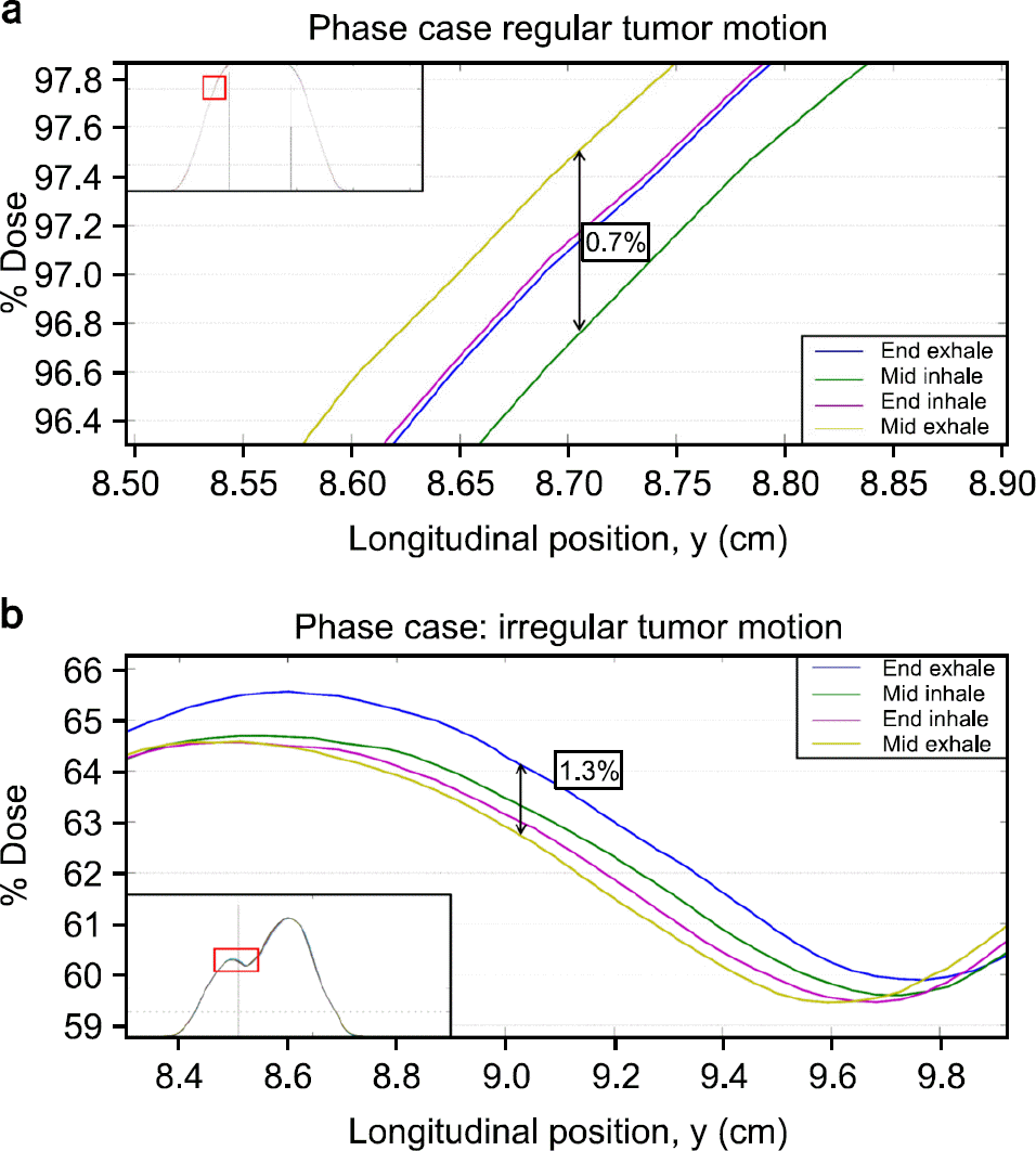 | Fig. 6.Dose profiles in the phase case (end inspiration, mid expiration, end expiration, and mid inspiration). The tumor motion was divided in (a) regular tumor motion case and (b) irregular tumor motion case. |
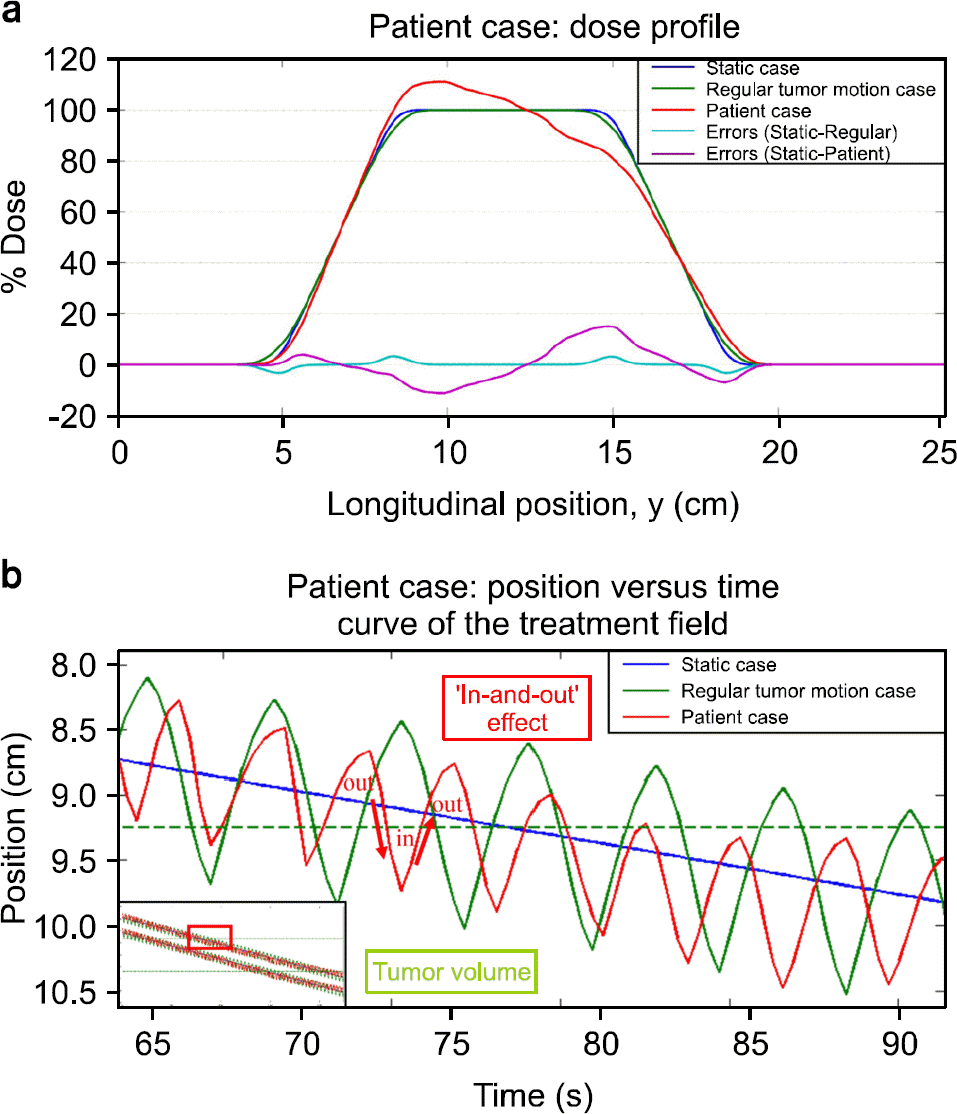 | Fig. 7.(a) Dose profiles in the patient case: no tumor motion case, regular tumor motion case, and real tumor motion case were compared. (b) Position versus time curve in the patient case. |
Table 1.
Experimental conditions used to acquire the static does.
Table 2.
Helical tomotherapy param information used in the simulation.
 XML Download