Abstract
In Tomotherapy the couch sags during the treatment due to the weight of the patient. In this study, we developed a simple method to obtain the amount of the sag and the pitch angle of the couch using the image processing technique of MVCT images in Tomotherapy. Using the method we evaluated the sag and pitch of couch for 22 head and neck patients and one craniospinal irradiation (CSI) patient. The sag and the average pitch angle of couch were 0.40∼1.54 mm and 0.7o for head and neck patients, respectively. For head and neck patients, the sag increased as the longitudinal length of the irradiation volume increased and the pitch angle showed no relationship with the longitudinal length. For the CSI patient the sag was 4.97 mm. Using the method the amount of the couch sag could be measured easily and the measured data could be useful in determination of margins considering the table sag error.
Go to : 
References
1. Ramsey C, Dube S, Hendee WR, et al. Is is necessary to validate each individual IMRT treatment plan before delivery Med Phys. 30:2271–2273. 2003.
2. Olivera GH, Shepard DM, Ruchala KJ, et al. “Tomotherapy.” In The Modern Technology of Radiation Oncology: A Compendium for Medical Physicists and Radiation Oncologists. (Madison, WI) 521–587 (. 1999.
3. Ruchala KJ, Olivera GH, Kapatoes J, et al. Limited-data image registration for radiotherapy positioning and verification. Int J Radiat Oncol Biol Phys. 54:592–605. 2002.

4. mackie TR, Olivera GH, Kapatoes JM, et al. Intensitymodulated radiation therapy. The state of the art. AAPM Summer School Proceedings. Colorado Springs, CO: Med Phys. Publishing 247–284 (. 2003.
5. Boswell SA, Jeraj R, Ruchala KJ, et al. A novel method to correct for pitch and yaw patient setup errors in helical tomotherapy. Med Phys. 32:1630–1639. 2005.

6. Logan DL. First course in the element method. (Boston: PWS Publishing Company, Inc).
7. Kazubek M. Wavelet domain image denoising by thresholding and Wiener filtering. Signal Processing Letters IEEE. 10:324–326. 2003.

8. Kaiser A, Schultheiss TE, Wong JYC, et al. Pitch, roll, and yaw variations in patient positioning. Int J Radiation Oncology Biol Phys. 66:949–995. 2006.

9. Hornick DC, Litzenberg DW, Lam KL, et al. A tilt and roll device for automated correction of rotational setup errors. Med Phys. 25:1739–1740. 1998.

10. Litzenberg DW, Balter JM, Hornic DC, et al. A mathematical model for correcting patient setup errors using a tilt and roll device Med Phys. 26:2586–2588. 1999.
Go to : 
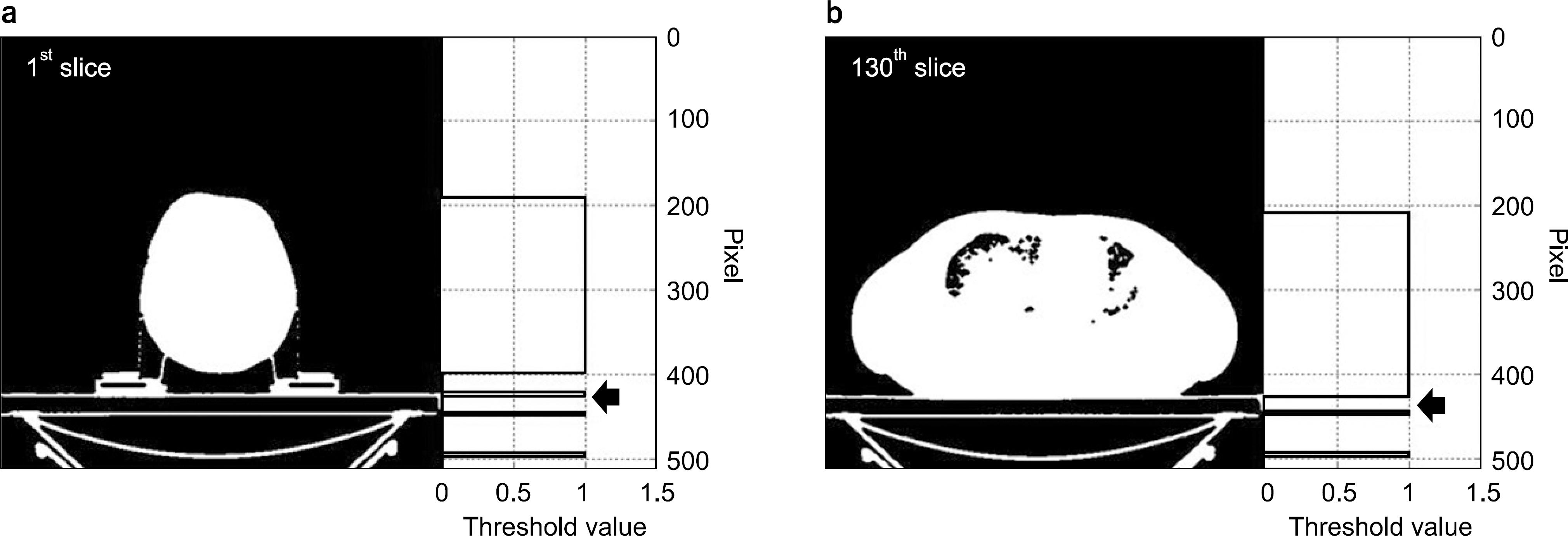
 | Fig. 2.방사선 단층영상의 영상 이진화 결과. (a) 단층영상의 첫 번째 슬라이스에서의 영상 이진화 결과와 영상 중앙에서 종축으로의 프로파일. (b) 단층영상의 마지막 슬라이스에서의 영상 이진화 결과와 영상 중앙에서 종축으로의 프로파일. |
 | Fig. 3.MVCT의 환자 테이블 처짐 분석 결과. (a) 치료범위에 따른 환자테이블 처짐. (b) 처짐 각도의 측정 결과. (c) 환자 체중에 따른 환자테이블의 처짐 각도. |


 PDF
PDF ePub
ePub Citation
 Print
Print


 XML Download