

 PDF
PDF ePub
ePub Citation
 Print
Print


Abstract
To see the discrepancies between the calculated and the delivered dose distribution of IMRT fields for respiratory-induced moving target according to the motion ranges. Four IMRT plans in which there are five fields, for lung and liver patients were selected. The gantry angles were set to 0° for every field and recalculated using TPS (Eclipse Ver 8.1, Varian Medical Systems, Inc., USA). The ion-chamber array detector (MatriXX, IBA Dosimetry, Germany) was placed on the respiratory simulating platform and made it to move with ranges of 1, 2, and 3 cm, respectively. The IMRT fields were delivered to the detector with 30∼70% gating windows. The comparison was performed by gamma index with tolerance of 3 mm and 3%. The average pass rate was 98.63% when there's no motion. When 1.0, 2.0, 3.0 cm motion ranges were simulated, the average pass rate were 98.59%, 97.82%, and 95.84%, respectively. Therefore, ITV margin should be increased or gating windows should be decreased for targets with large motion ranges.
Go to : 
References
1. Ezzell GA, Burmeister JW, Dogan N, et al. IMRT commissioning: Multiple institution planning and dosimetry comparisons, a report from AAPM Task Group 119, Med Phys. 36:5359–5373. 2009.
2. Ahn WS, Cho BC. Intensity modulated radiation therapy commissioning and quality assurance: Implementation of AAPM TG119. Progress in Medical Physics. 22:99–105. 2011.
3. Hussein M, Rowshanfarzad P, Ebert MA, Nisbet A, Clark CH. A comparison of the gamma index analysis in various commercial IMRT/VMAT QA systems. Radiother Oncol. 109:370–376. 2013.

4. Arumugam S, Xing A, Goozee G, Holloway L. Independent calculation-based verification of IMRT plans using a 3D dose-calculation engine. Med Dosim. 38:376–384. 2013.
5. Anjum MN, Parker W, Ruo R, Aldahlawi I, Afzal M. IMRT quality assurance using a second treatment planning system. Med Dosim. 35:274–279. 2010.
6. Li G, Zhang Y, Jiang X, et al. Evaluation of the ArcCHECK QA system for IMRT and VMAT verification. Physica Medica. 29:295–303. 2013.
7. Guillot M, Gingras L, Archambault L, Beddar S, Beaulieu L. Performance assessment of a 2D array of plastic scintillation detectors for IMRT quality assurance. Phys Med Biol. 58:4439–4454. 2013.
8. Sabet M, Rowshanfarzad P, Vial P, Menk FW, Greer PB. Transit dosimetry in IMRT with an a-Si EPID in direct detection configuration. Phys Med Biol. 57:N295–N306. 2012.
9. Vial P, Gustafsson H, Oliver L, Baldock C, Greer PB. Direct-detection EPID dosimetry: investigation of a potential clinical configuration for IMRT verification. Phys Med Biol. 54:7151–7169. 2009.
10. Carlone M, Cruje C, Rangel A, et al. ROC analysis in patient specific quality assurance. Med Phys. 40:0421031–0421037. 2013.
11. Intensity Modulated Radiation Therapy Collaborative Working Group: Intensity modulated radiotherapy: current status and issues of interest. Int J Radiat Oncol Biol Phys. 51:880–917. 2001.
12. Wilcox EE, Daskalov GM, Pavlonnis III, et al. Dosimetric verification of intensity modulated radiation therapy of 172 patients treated for various disease sites: comparison of EBT film dosimetry, ion chamber measurements, and independent MU Calculations. Med Dosim. 33:303–309. 2008.
13. Anjum MN, Parker W, Ruo R, Afzal M. Evaluation criteria for film based intensity modulated radiation therapy quality assurance. Physica Medica. 26:38–43. 2010.
14. Roesink JM, Moerland MA, Battermann JJ, Hordijk GJ, Terhaard CHJ. Qantitative dose-volume response analysis of changes in parotid gland function after radiotheraphy in the head-and-neck region. Int J Radiat Oncol Biol Phys. 51:938–946. 2001.
15. Sasaki R, Soejima T, Matsumoto A, et al. Clinical significance of serum pulmonary surfactant proteins A and D for the early detection of radiation pneumonitis. Int J Radiat Oncol Biol Phys. 50:301–307. 2001.
16. Lee N, Xia P, Quivey JM, et al. Intensity-modulated radiotherapy in the treatment of nasopharyngeal carcinoma: an update of the UCSF experience. Int J Radiat Oncol Biol Phys. 53:12–22. 2002.
17. Nutting CM, Convery JD, Cosgrove PV, et al. Reduction of small and large bowel irradiation using an optimized intensitymodulated pelvic radiotherapy technique in patients with prostate cancer. Int J Radiat Oncol Biol Phys. 48:649–656. 2000.
18. Sanguineti G, Cavey ML, Endres EJ, et al. Is IMRT needed to spare the rectum when pelvic lymph nodes are part of the initial treatment volume for prostate cancer? Int J Radiat Oncol Biol Phys. 64:151–160. 2006.
19. Luxton G, Hancock SL, Boyer AL. Dosimetry and radiobiologic model comparison of IMRT and 3D conformal radiotherapy in treatment of carcinoma of the prostate. Int J Radiat Oncol Biol Phys. 59:267–284. 2004.
20. Wang-Chesebro A, Xia P, Coleman J, Akazawa C, Roach III. Intensity-modulated radiotherapy improves lymph node coverage and dose to critical structures compared with three-dimensional conformal radiation therapy in clinically localized prostate cancer. Int J Radiat Oncol Biol Phys. 66:654–662. 2006.
21. Wang L, Hoban P, Paskalev K, et al. Dosimetric advantage and clinical implication of a micro-multileaf collimator in the treatment of prostate with intensitymodulated radiotherapy. Med Dosim. 30:97–103. 2005.
22. ICRU REPORT 62: Prescribing, Recording and Reporting Photon Beam Therapy (Supplement to ICRU Report 50). International Commission in Radiation Units and Measurements (. 1999.
23. Chu SS, Cho KH, Lee CG, Suh CO. Development of conformal radiotherapy with respiratory gate device. Radiat Oncol J. 20:41–52. 2002.
24. Kini VR, Vedam SS, Keall PJ, et al. Patient training in respiratory-gated radiotherapy. Med Dosim. 28:7–11. 2003.
25. Lim S, Park SH, Ahn SD, et al. Guiding curve based on the normal breathing as monitored by thermocouple for regular breathing. Med Phys. 34:4514–4518. 2007.
26. Lim S. Research on the respiratory synchronization system for dynamic tumor tracking radiation therapy. Gyeonggi, Kyonggi University doctorate thesis (. 2008.
27. Vedam SS, Kini VR, Keall PJ, et al. Quantifying the predictability of diaphragm motion during respiration with a noninvasive external marker. Med Phys. 30(4):505–513. 2003.
28. Cervino LI, Du J, Jiang SB. MRI-guided tumor tracking in lung cancer radiotherapy. Phys Med Biol. 56:3773–3785. 2011.
29. IBA Dosimetry: I'mRT MatriXX: The new standard in 2D IMRT pre-treatment verification. http://www.iba-dosimetry.com.
30. Ma SY, Choi JH, Jeung TS, Lim S. Quantitative evaluation of gated radiation therapy using gamma index analysis. Progress in Medical Physics. 24:198–203. 2013.
31. Lim S, Ahn SD, Park SH, et al. Study of respiration simulating phantom using thermocouple-based respiration monitoring mask. Radiat Oncol J. 23:217–222. 2005.
32. Yoon GJ, Kwak JW, Cho BC, et al. Development of new 4D phantom model in respiratory gated volumetric modulated arc therapy for lung SBRT. Progress in Medical Physics. 25:100–109. 2014.
Go to :
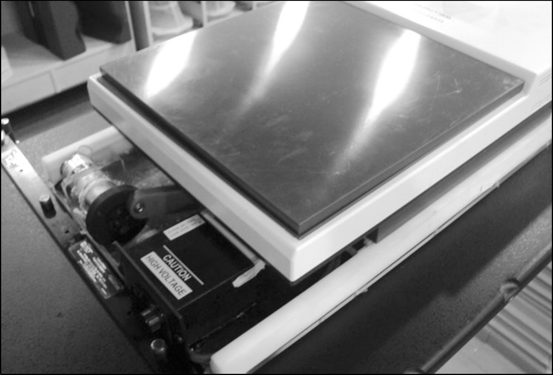 | Fig. 1.To simulate the patients' respiratory motion, the moving platform was fabricated in laboratory. The solid water phantom with thickness of 1.3 cm and the MatriXX were placed on the simulating platform as shown figure. The SSD was 100 cm and the depth of measurement layer became 1.6 cm, which Dmax for 6 MV photon beams. We let the platform move with range of 0, 1, 2, and 3 cm and with cycle of 3.6 s. |
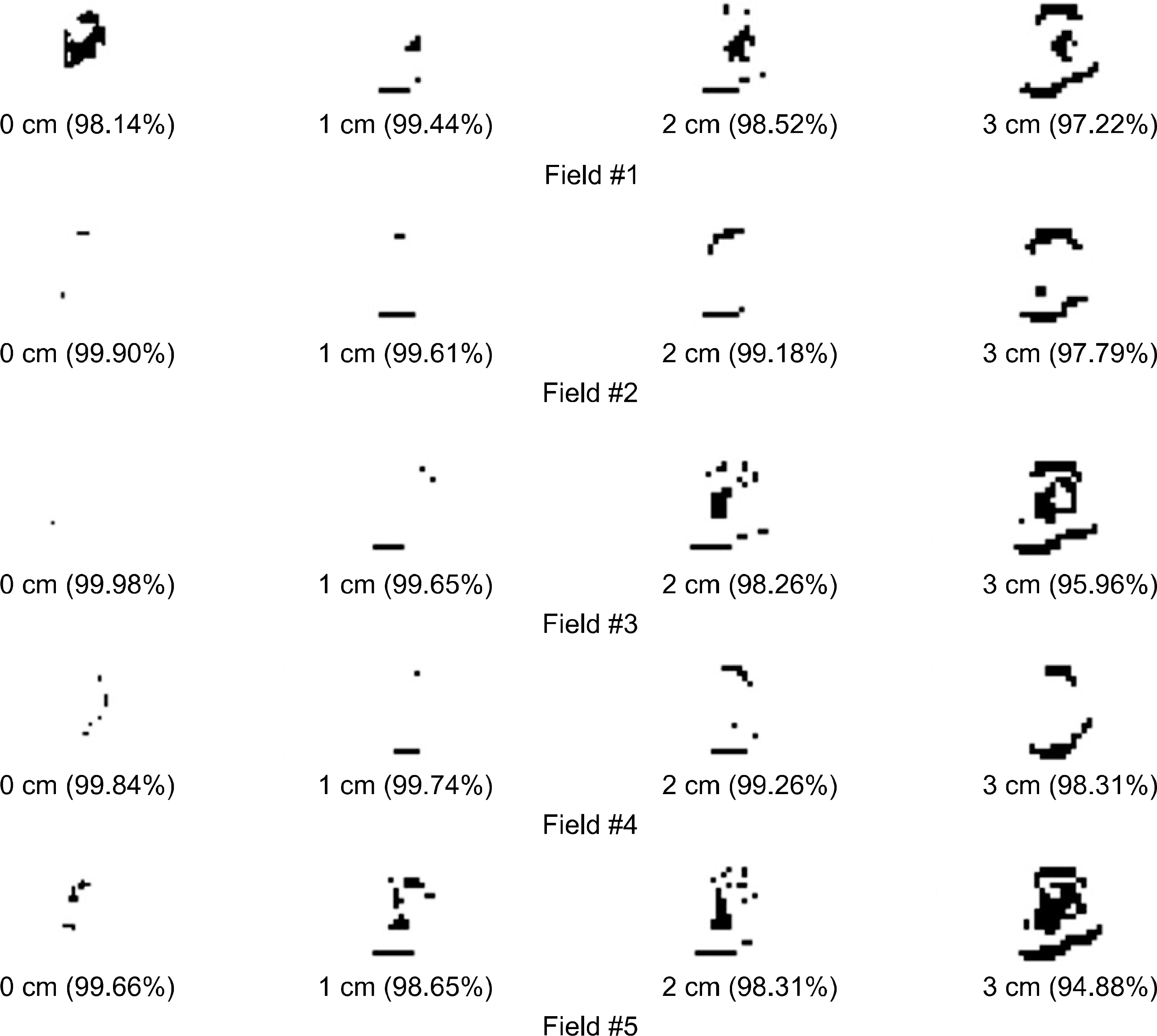 | Fig. 2.Example of five respiratory gated IMRT fields for case of A patient; the phantom simulated respiratory motion with four scenario (0, 1, 2, and 3 cm motion range and 3.6 s cycle). Gamma index were used with 3 mm/3% tolerance to compare calculated and measured dose distribution. White and black area in the figures represent pass and fail respectively. |
Table 1.
Respiratory patterns for 48 lung and liver cancer patients (SD: Standard deviation).
Table 2.
The pass rate according to the motion ranges: The pass rate for the calculated and the measured dose distribution for the gated IMRT according to the target motion ranges; The pass rates tend to decrease with the motion range.
 XML Download