

 PDF
PDF ePub
ePub Citation
 Print
Print


Abstract
The aim of this study is to develop a new software tool for 3D dose verification using PRESAGEREU Gel dosimeter. The tool included following functions: importing 3D doses from treatment planning systems (TPS), importing 3D optical density (OD), converting ODs to doses, 3D registration between two volumetric data by translational and rotational transformations, and evaluation with 3D gamma index. To acquire correlation between ODs and doses, CT images of a PRESAGEREU Gel with cylindrical shape was acquired, and a volumetric modulated arc therapy (VMAT) plan was designed to give radiation doses from 1 Gy to 6 Gy to six disk-shaped virtual targets along z-axis. After the VMAT plan was delivered to the targets, 3D OD data were reconstructed from 512 projection data from VistaTM optical CT scanner (Modus Medical Devices Inc, Canada) per every 2 hours after irradiation. A curve for converting ODs to doses was derived by comparing TPS dose profile to OD profile along z-axis, and the 3D OD data were converted to the absorbed doses using the curve. Supra-linearity was observed between doses and ODs, and the ODs were decayed about 60% per 24 hours depending on their magnitudes. Measured doses from the PRESAGEREU Gel were well agreed with the TPS doses at central region, but large under-doses were observed at peripheral region at the cylindrical geometry. Gamma passing rate for 3D doses was 70.36% under the gamma criteria of 3% of dose difference and 3 mm of distance to agreement. The low passing rate was resulted from the mismatching of the refractive index between the PRESAGE gel and oil bath in the optical CT scanner. In conclusion, the developed software was useful for 3D dose verification from PRESAGE gel dosimetry, but further improvement of the Gel dosimetry system were required.
Go to : 
References
1. Fraass B, Doppke K, Hunt M, et al. American Association of Physicists in Medicine Radiation Therapy Committee Task Group 53: Quality assurance for clinical radiotherapy treatment planning. Med. Phys. 25(10):1773–1829. 1998.

2. Schreibmann E, Dhabaan A, Elder E, Fox T. Patientspecific quality assurance method for VMAT treatment delivery. Med. Phys. 36(10):4530–4535. 2009.
3. Zhen H, Benjamin EN, Wolfgang A, Tomé: Moving from gamma passing rates to patient DVH-based QA metrics in pretreatment dose QA. Med. Phys. 38(10):5477–5489. 2011.
4. Benjamin EN, Zhen H, Wolfgang A, Tomé: Per-beam, planar IMRT QA passing rates do not predict clinically relevant patient dose errors. Med. Phys. 38(2):1037–1044. 2011.
5. Wu C, Kelly E. Hosier, Kristen E. Beck, et al: On using 3D γ-analysis for IMRT and VMAT pretreatment plan QA. Med. Phys. 39(6):3051–3059. 2012.
6. Carrasco P, Jornet N, Latorre A, Eudaldo T, Ruiz A, Ribas M. 3D DVH-based metric analysis versus per-beam planar analysis in IMRT pretreatment verification. Med. Phys. 39(8):5040–5049. 2012.
7. Visser R, Wauben DJL, Groot M, et al. Efficient and reliable 3D dose quality assurance for IMRT by combining independent dose calculations with measurements. Med. Phys. 40(2):021710–1. -6 (. 2013.
8. Fredh A, Scherman JB, Fog LS, Rosenschöld PM. Patient QA systems for rotational radiation therapy: A comparative experimental study with intentional errors. Med. Phys. 40:031716. 2013.
9. Juang T, Newton J, Niebanck M, Benning R, Adamovics J, Oldham M. Customising PRESAGE® for diverse applications. Journal of Physics: Conference Series. 444:1–5. 2013.
10. Qian X, Adamovics J, Wuu C. Performance of an improved first generation optical CT scanner for 3D dosimetry. Phys. Med. Biol. 58:N321–N331. 2013.
11. Juang T, Grant R, Adamovics J, Ibbott G, Oldham M. On the feasibility of comprehensive high-resolution 3D remote dosimetry. Med. Phys. 41(7):071706–1. -11 (. 2014.
12. Cho SJ, Chung YL, Lee SH, et al. A Study of Optimized MRI Parameters for Polymer Gel Dosimetry, Korean J Med Phys. 23(2):71–80. 2012.
13. Jang JS, Kwon SI. 3-Dimensional Dosimetry of Small Field Photon Beam, Korean J Med Phys. 23(1):54–61. 2012.
14. Lee KN, Lee DJ, Suh TS. 3-Dimensional Verification Technique for Target Point Error, Korean J Med Phys. 22(1):35–41. 2011.
15. Jung JY, Lee CI, Min JH, Kim YL, Lee SY, Suh TS. A Study on Dose Response of MAGAT (Methacrylic Acid, Gelatin Gel and THPC) Polymer Gel Dosimeter Using X-ray CT Scanner. Korean J Med Phys. 21(1):1–8. 2010.
16. Lopatiuk-Tirpak O, Langen KM, Meeks SL, Kupelian PA, Zeidan OA, Maryanski MJ. Performance evaluation of an improved optical computed tomography polymer gel dosimeter system for 3D dose verification of static and dynamic phantom deliveries, Med. Phys. 35(9):3847–3859. 2008.
17. Jirasek A. Experimental investigations of polymer gel dosimeters, Journal of Physics: Conference Series. 56:23–34. 2006.
18. Jordan K. Review of recent advances in radiochromic materials for 3D dosimetry, Journal of Physics: Conference Series. 250:1–7. 2010.
19. Pierquet M, Thomas A, Adamovics J, Oldham M. An investigation into a new re-useable 3D radiochromic dosimetry material, PresageREU, Journal of Physics: Conference Series. 250:1–4. 2010.
20. Adamovics J, Maryanski MJ. CHARACTERISATION OF PRESAGETM: A NEW 3-D RADIOCHROMIC SOLID POLYMER DOSEMETER FOR IONISING RADIATION, Radiation Protection Dosimetry, 120 (1–4):. 107–112. (. 2006.
21. Doran SJ, Krstajic N, Adamovics J, Jenneson PM. Optical CT scanning of PRESAGETM polyurethane samples with a CCD-based readout system, Journal of Physics: Conference Series. 3:240–243. 2004.
Go to :
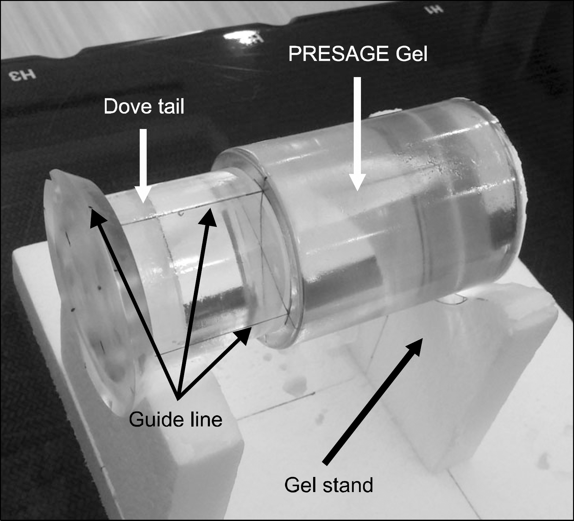 | Fig. 1.Setup of PRESAGEREU gel dosimeter: Guide lines were drawn on the dose tail only rather than to gel body. |
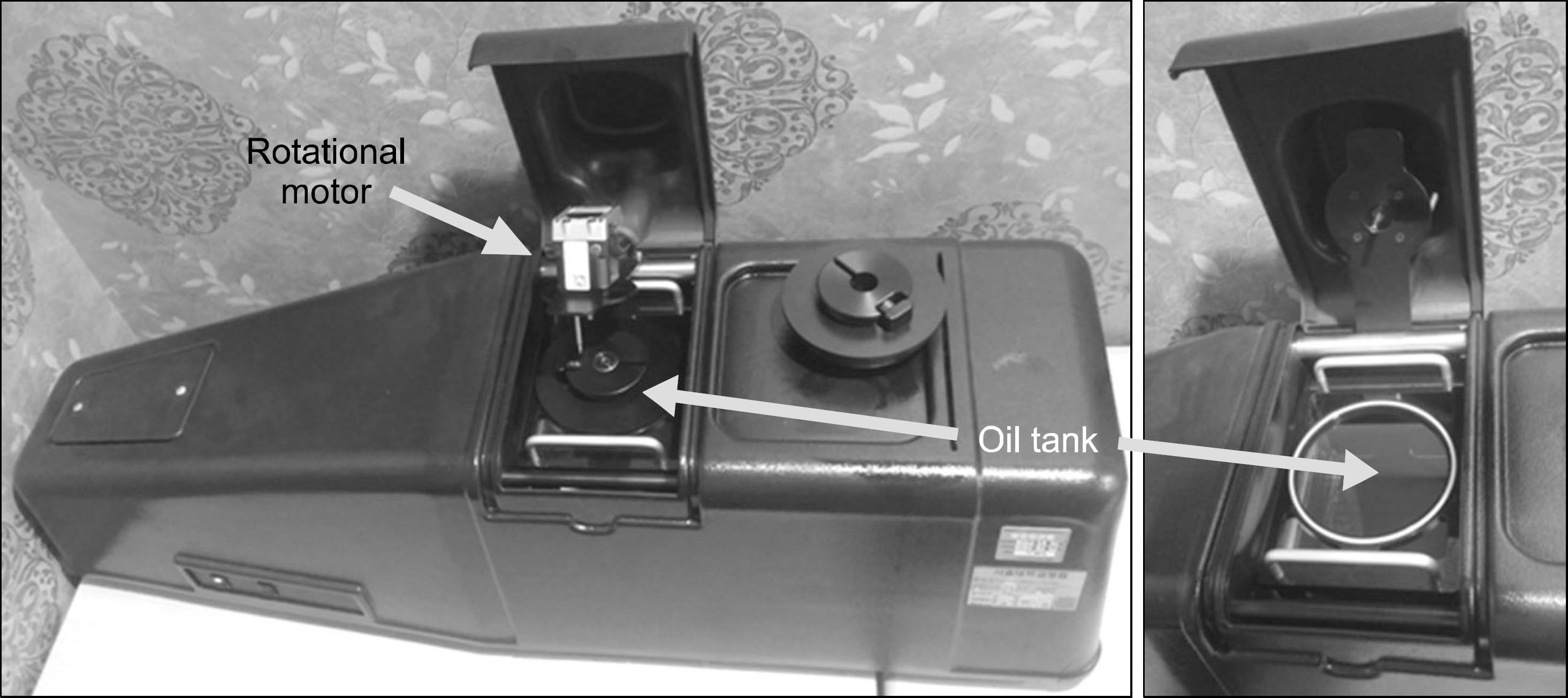 | Fig. 2.VistaTM optical CT scanner. dove tail on the gel can be attached to the rotational motor, and gel body is immersed into oil tank. |
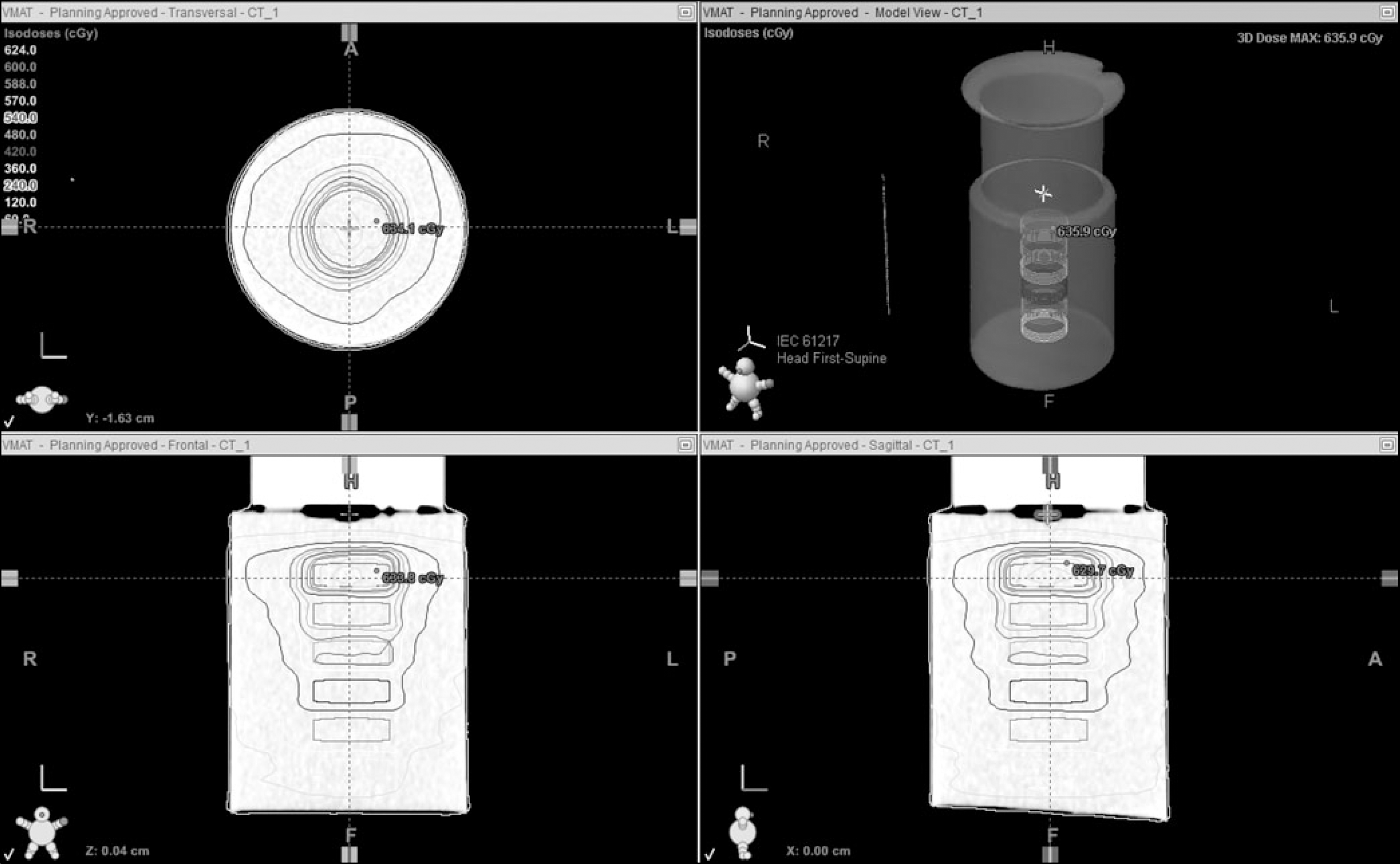 | Fig. 3.Radiation dose distributions in PRESAGE gel at the treatment planning system. Six virtual treatment targets were delineated along the cylinder axis, and absorbed doses from 1 Gy to 6 Gy was delivered to each targets. |
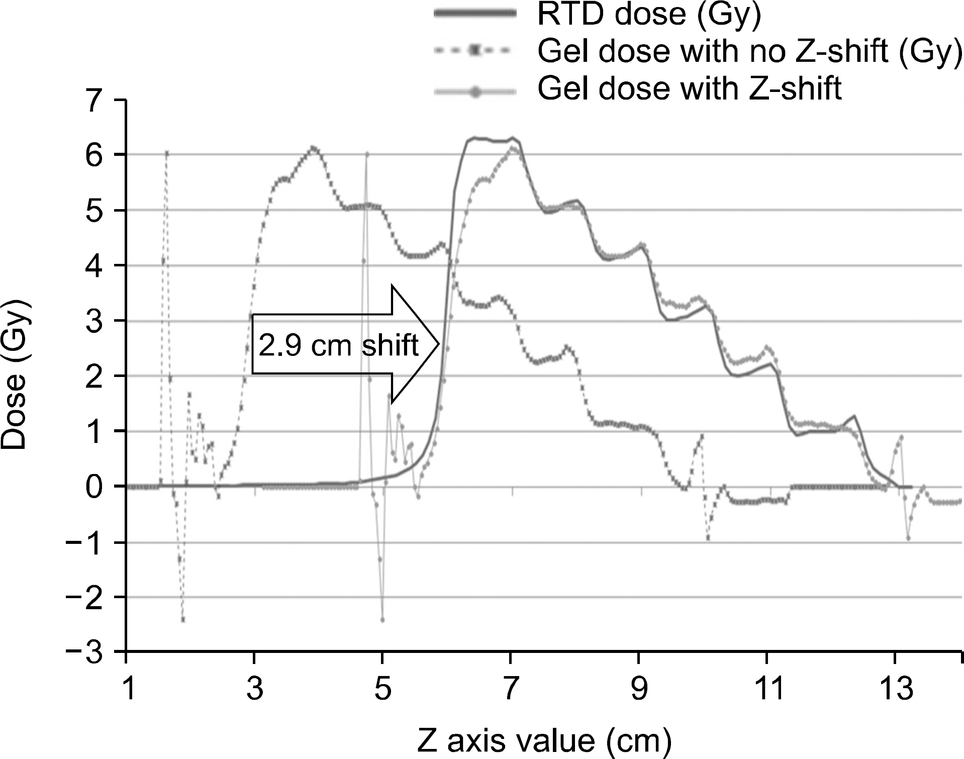 | Fig. 4.Dose profiles along cylindrical axis from TPS doses and PRESAGE gel doses. The PRESAGE gel doses were calculated by applying a single OD to Dose converting factor. Proper shift along z-axis of the gel dose make two profiles correctly matched. |
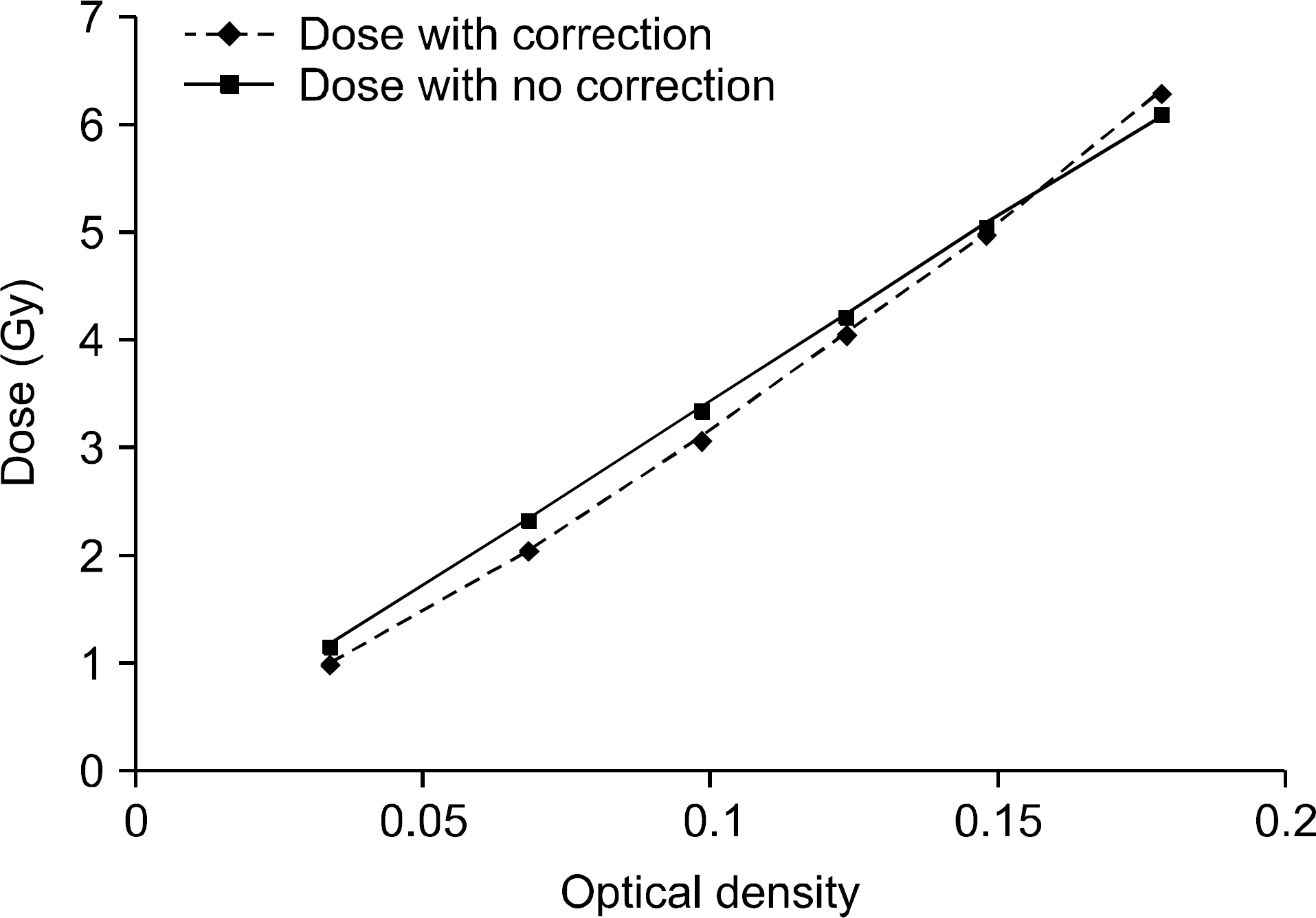 | Fig. 5.The correlation between optical density (OD) and absorbed doses. The curve of “Dose with correction” means that variable OD to dose converting factor is used to calculated doses from OD. The curve of “Dose without correction” used only a single OD to dose converting factor. |
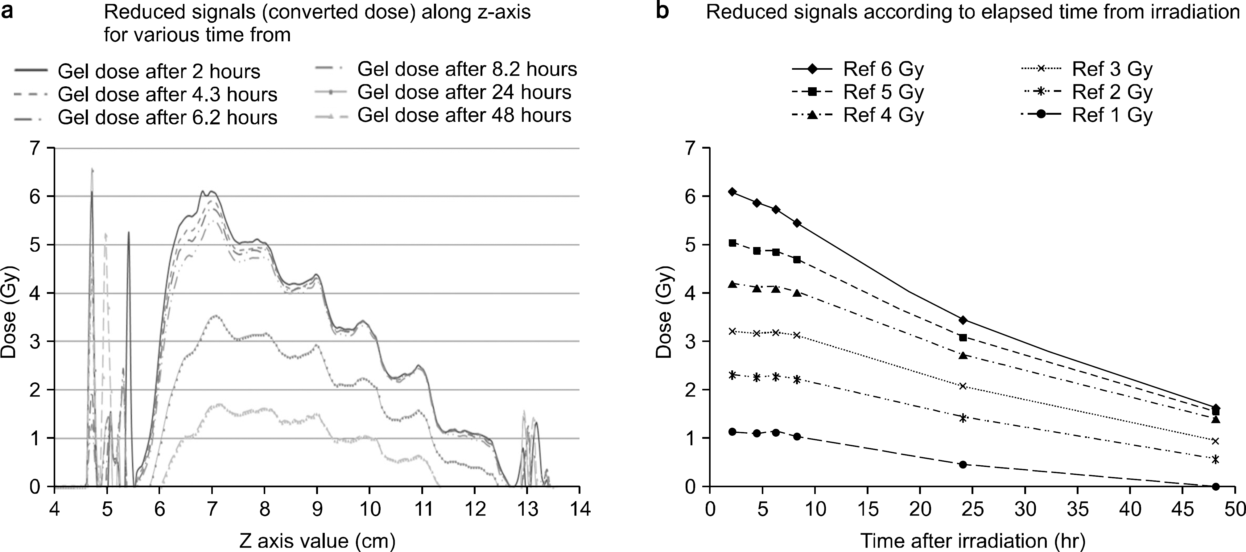 | Fig. 6.Decay of the optical densities (or converted doses) along the time after irradiation. |
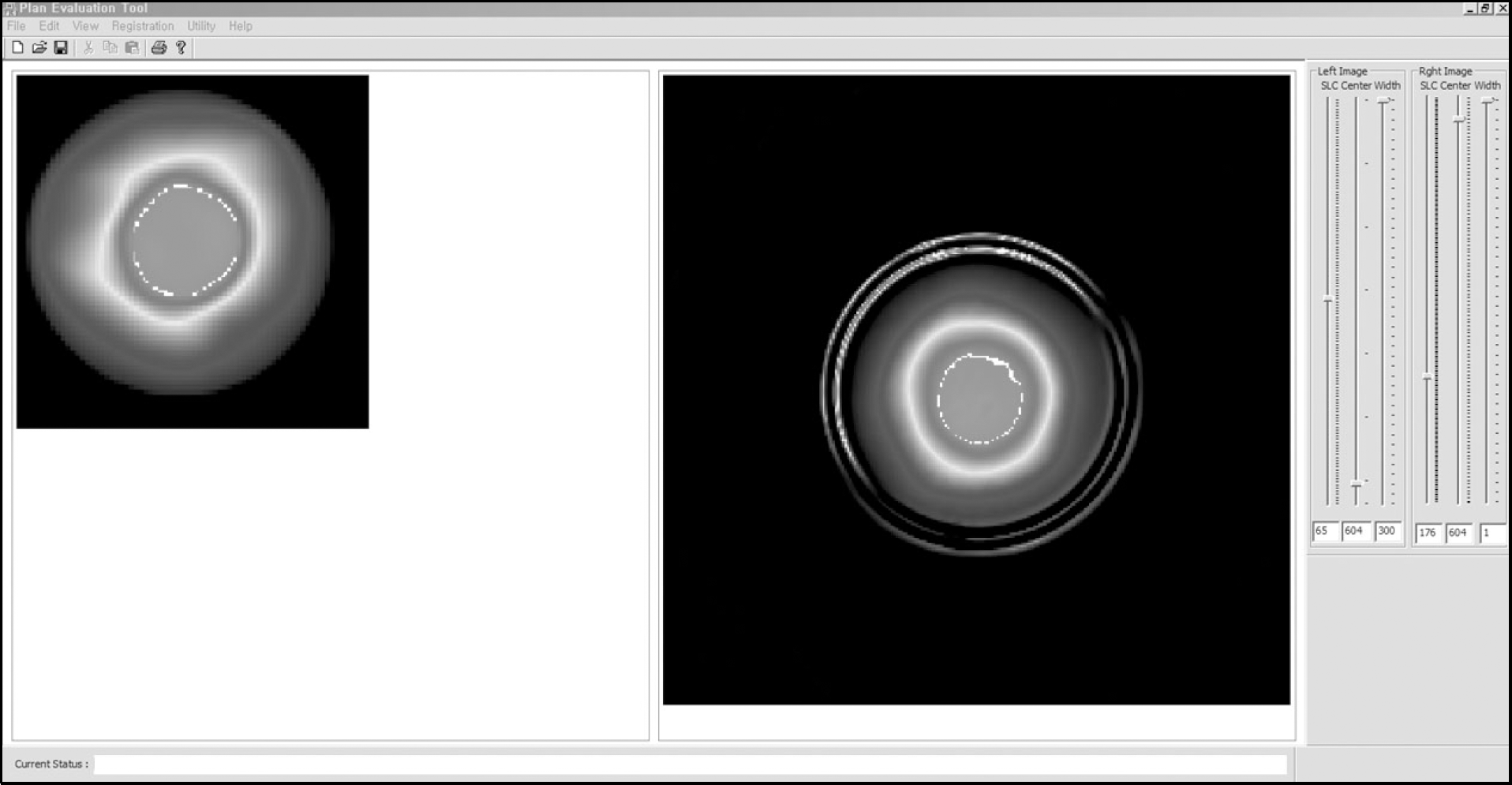 | Fig. 7.Graphic User Interface of the developed software tool: left window shows calculated dose distribution from TPS, right window shows measured dose distributions from gel dosimeter. Slide bars at right side can control the current slice of the 3D volume, minimum and maximum threshold of the dose normalization. |
 | Fig. 8.The tool for the alignment of 3D gel dose to the TPS doses and evaluation of tow dose distributions: (a) before alignment (b) after alignment by x, y, z translational and rotational transformation. There are some interfaces for ① x, y, z offset [mm] ② rotational offset [degree], ③ Display mode option ④ Passing rates from 3D Gamma index method ⑤ Criteria of the gamma index method. |
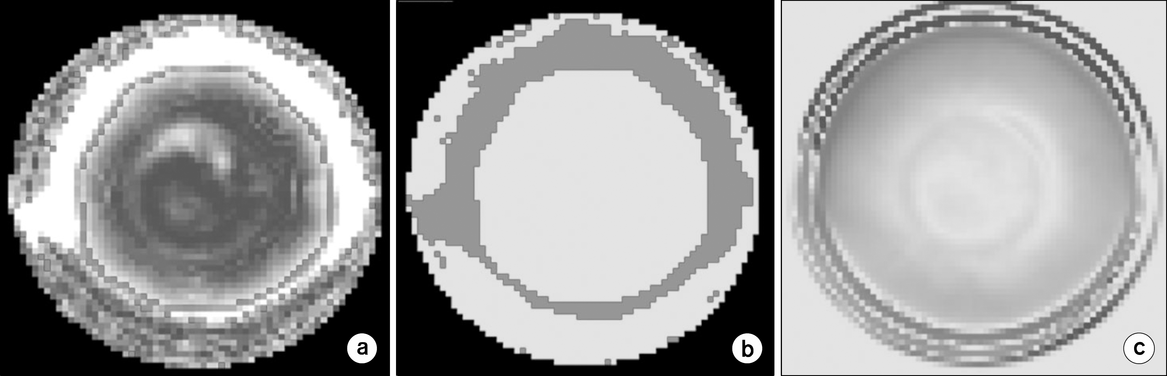 | Fig. 9.Example of the comparison between TPS doses and gel doses: (a) Gamma index map, (b) Gamma pass map, (c) dose difference map. |
Table 1.
Optical density, expected dose from treatment planning system, measured absorbed dose using single OD to dose converting factor.
 XML Download