Abstract
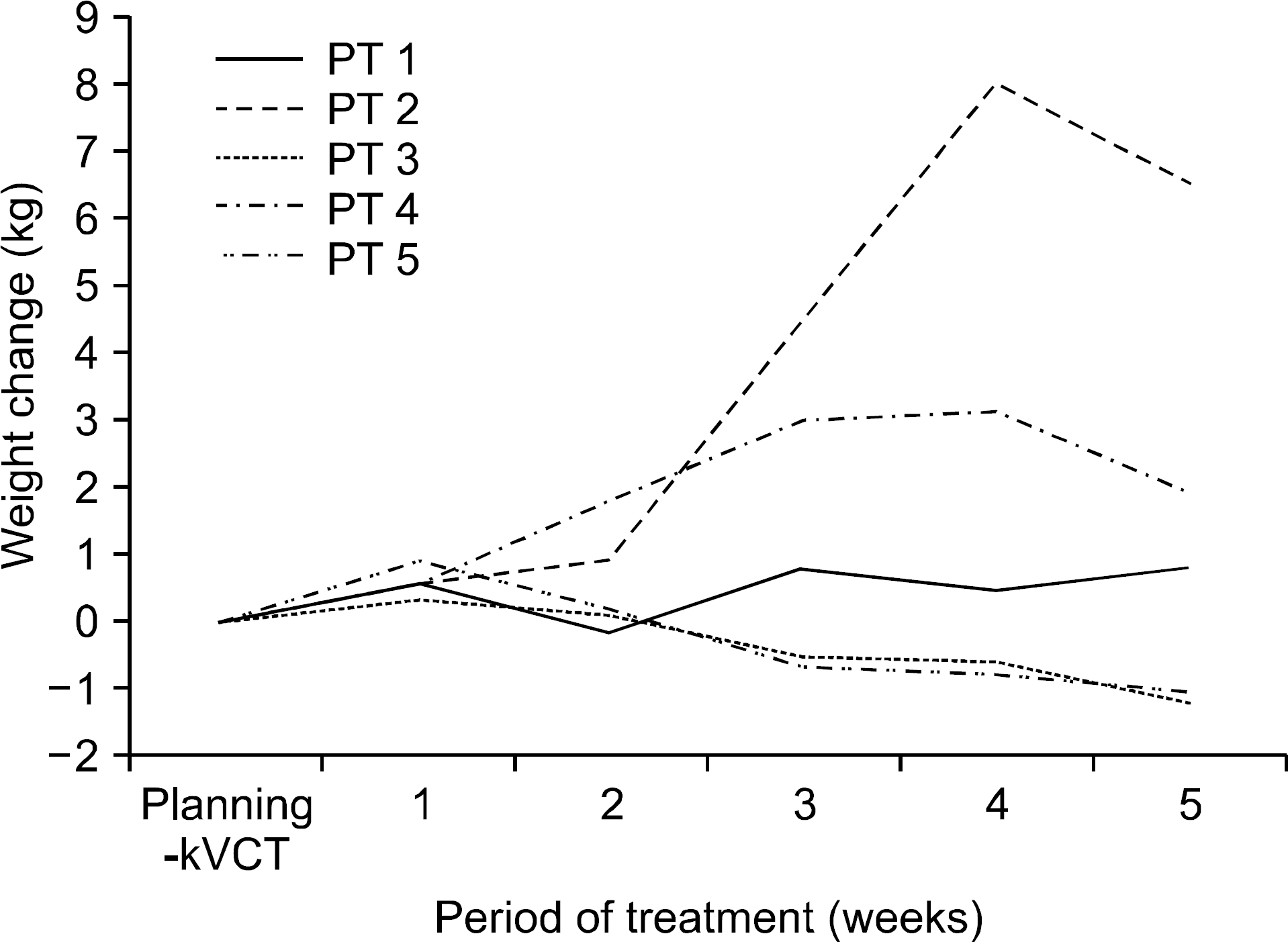
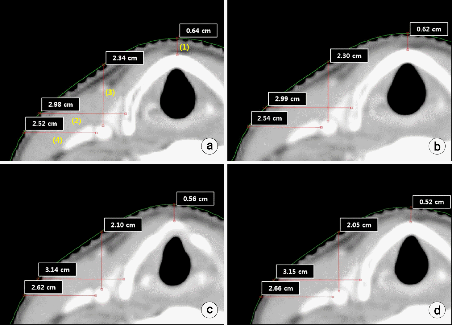
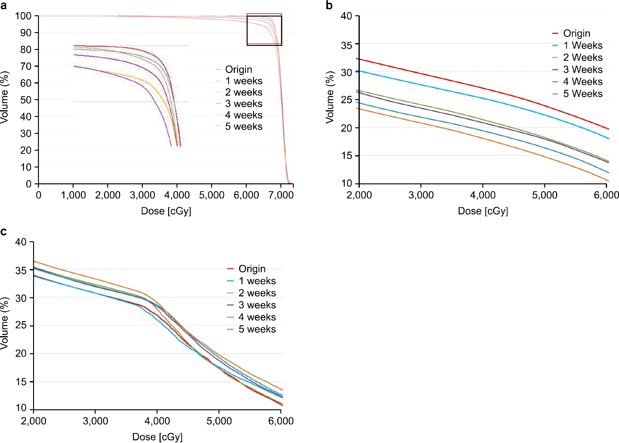
The purpose of this study is to evaluate the variation of the dose which is delivered to the patients with glottis cancer under IMRT (intensity modulated radiation therapy) by using the 3D registration with CBCT (cone beam CT) images and the DIR (deformable image registration) techniques. The CBCT images which were obtained at a one-week interval were reconstructed by using B-spline algorithm in DIR system, and doses were recalculated based on the newly obtained CBCT images. The dose distributions to the tumor and the critical organs were compared with reference. For the change of volume depending on weight at 3 to 5 weeks, there was increased of 1.38∼2.04 kg on average. For the body surface depending on weight, there was decreased of 2.1 mm. The dose with transmitted to the carotid since three weeks was increased compared be more than 8.76% planned, and the thyroid gland was decreased to 26.4%. For the physical evaluation factors of the tumor, PITV, TCI, rDHI, mDHI, and CN were decreased to 4.32%, 5.78%, 44.54%, 12.32%, and 7.11%, respectively. Moreover, Dmax, Dmean, V67.50, and D95 for PTV were increased or decreased to 2.99%, 1.52%, 5.78%, and 11.94%, respectively. Although there was no change of volume depending on weight, the change of body types occurred, and IMRT with the narrow composure margin sensitively responded to such a changing. For the glottis IMRT, the patient's weight changes should be observed and recorded to evaluate the actual dose distribution by using the DIR techniques, and more the adaptive treatment planning during the treatment course is needed to deliver the accurate dose to the patients.
Go to : 
References
1. Rosenthal DI, Fuller CD, Barker JL Jr, Mason B, Garcia JA, Lewin JS. Simple carotid-sparing intensitymodulated radiotherapy technique and preliminary experience for T1–2 glottic cancer. Int J Radiat Oncol Biol Phys. 77(2):455–461. 2010.

2. Geets X, Tomsej M, Lee JA, et al. Adaptive biological imageguided IMRT with anatomic and functional imaging in pharyngolaryngeal tumors: Impact on target volume delineation and dose distribution using helical tomotherapy. Radiother Oncol. 85:105–115. 2007.

3. Wu Q, Chi Y, Chen PY, et al. Adaptive replanning strategies accounting for shrinkage in head and neck IMRT. Int J Radiat Oncol Biol Phys. 75:924–932. 2009.

4. Hunter KU, Fernandes LL, Vineberg KA, et al. Parotid glands dose-effect relationships based on their actually delivered doses: implications for adaptive replanning in radiation therapy of head-and-neck cancer. Int J Radiat Oncol Biol Phys. 87(4):676–682. 2013.

5. Nishimura Y, Nakamatsu K, Shibata T, et al. Importance of the initial volume of parotid glands in xerostomia for patients with head and neck cancers treated with IMRT. Jpn J Clin Oncol. 35:375–379. 2005.

6. Schwartz DL, Garden AS, Thomas J, et al. Adaptive radiotherapy for head-and-neck cancer: Initial clinical outcomes from a prospective trial, Int. J. Radiat. Oncol. Biol. Phys. 83:986–993. 2011.
7. Mencarelli A, van Beek S, van Kranen S, Rasch C, van Herk M, Sonke JJ. Validation of deformable registration inhead and neck cancer using analysis of variance. Med Phys. 39(11):6879–6884. 2012.
8. Yan D, Liang J. Expected treatment dose construction and adaptive inverse planning optimization: implementation for offline head and neck cancer adaptive radiotherapy. Med Phys. 40(2):021719. 2013.

9. Mike O, Jeff C, Eugene W, Jake VD, Francisco P. A treatment planning study comparing whole breast radiation therapy against conformal, IMRT and tomotherapy for accelerated partial breast Irradiation. Radiother Oncol. 82:317–323. 2007.
10. Bhide SA, Davies M, Burke K, et al. Weekly volume and dosimetric changes during chemoradiotherapy with intensitymoudulated radiation therapy for head and neck cancer: a prospective observational study. Int. J. Radiat. Oncol. Biol. Phys. 76:1360–1368. 2010.
11. Barker JL, Gargen AS, Ang KK, et al. Quantification of volumetric and geometric changes occurring during fractionated radiotherapy for head-and-neck cancer using an integrated CT/linear accelerator system, Int. J. Radiat. Oncol. Biol. Phys. 59:960–970. 2004.
12. Schwartz DL, Garden AS, Thomas J, et al. Adaptive radiotherapy for head-and-neck cancer: Initial clinical outcomes from a prospective trial, Int. J. Radiat. Oncol. Biol. Phys. 83:986–993. 2011.
13. Lu W, Chen ML, Olivera GH, et al. Fast free-form deformable registration via calculus of variation. Phys. Med. Biol. 49:3067–3087. 2004.
14. Lu W, Olivera GH, Chen Q, et al. Deformable registration of the planning image(KVCT) and the daily images(MVCT) for adaptive radiation therapy. Phys. Med. biol. 51:4357–4374. 2006.
15. Ahn PH, Chen CC, Ahn AI, et al. Adaptive planning intensitymodulated radiation therapy for head and neck cancers: single-institution experience and clinical implications. Int. J. Radiat. Oncol. Biol. Phys. 3:677–685. 2011.
16. Feigenberg SJ, Lango M, Nicolaou N, Ridge JA. Intensitymodulated radiotherapy for early larynx cancer: is there a role? Int J Radiat Oncol Biol Phys 1. 68(1):2–3. 2007.

17. Martin JD, Buckley AR, Graeb D, Walman B, Salvian A, Hay JH. Carotid artery stenosis in asymptomatic patients who have received unilateral head-and-neck irradiation. Int J Radiat Oncol Biol Phys. 63:1197–1205. 2005.

18. Akgun Z, Atasoy BM, Ozen Z, et al. V30 as a predictor for radiation-induced hypothyroidism: a dosimetric analysis in patients who received radiotherapy to the neck. Radiation Oncology 9:. 104:1–5. 2014.

19. Cheng SW, Ting AC, Ho P, Wu LL. Accelerated progression of carotid stenosis in patients with previous external neck irradiation. J Vasc Surg. 39:409–415. 2004.

Go to : 
 | Fig. 1.The change of weight during the period of treatment. The difference of weight change for 1∼5 weeks after commencing the treatment based on the weight when the planning-kVCT has been acquired for the treatment plan. |
 | Fig. 2.Measurement and comparison of the distances between skin surface and each 4 points (OARs) in a typical patient. The change of body type occurs owing to the changing weight according to the progress of treatment. (a) planning-kVCT, (b) 1week, (c) 3week, (d) 5 week. (1) The distance between the anterior surface of thyroid cartilage and the skin. (2) The distance between the lateral surface of thyroid cartilage and the skin. (3) The distance between the anterior surface of carotid artery and the skin. (4) The distance between the lateral surface of carotid artery and the skin. |
 | Fig. 3.Comparison of dose volume histogram (DVHs) for different plans (original Vs. recalculation) from experimental data in a typical patient. (a) PTV, (b) thyroid gland, (c) carotid artery. |
Table 1.
Average of volume change on PTV and OARs.
Table 2.
Dose evaluation of PTV during the course of treatment.
Note: V67.50=the volume included in 67.50 Gy, D95=the dose included in 95% volume. PITV=PIV/PTV (PIV is the prescription isodose volume coverage for the target and normal tissues. PITV>1 and PITV<1 refers to the over treatment and under treatment regions, repectively.), TCI=PTVpd/PTV (PTVpd is the PTV coverage at PD, and PTV has usual meaning), rDHI=Dmin/Dmax, mDHI=D95/D5, CN=TCI/TR (CN accounts for the relative measurement of dosimetric target coverage and sparing of normal tissues in a treatment plan).
Table 3.
Dose evaluation of carotid artery.
Table 4.
Dose evaluation of thyroid gland.


 PDF
PDF ePub
ePub Citation
 Print
Print


 XML Download