

 PDF
PDF ePub
ePub Citation
 Print
Print


Abstract
In general radiotherapy, mega-voltage (MV) x-ray images are widely used as the unique method to verify radio-therapeutic fields. But, the image quality of MV images is much lower than that of kilo-voltage x-ray images due to scatter interactions. Since 1990s, studies for the scatter correction have performed with digital-based MV imaging systems. In this study, a novel method for the scatter correction is suggested using scatter to primary ratio (SPR), instead of conventional methods such as digital image processing or scatter kernel calculations. We measured two MV images with and without a solid water phantom describing a patient body with given imaging conditions, and calculated un-attenuated ratios. Then, we obtained SPR distributions for the scatter correction. For experimental validation, a line-pair (LP) phantom using several Al bars and a clinical pelvis MV image was used. As the result, scatter signals of the LP phantom image were successfully reduced so that original density distribution of the phantom was restored. Moreover, image contrast values increased after SPR correction at all ROIs of the clinical image. The mean value of increases was 48%. The SPR correction method suggested in this study has high reliability because it is based on actually measured data. Also, this method can be easily adopted in clinics without additional cost. We expected that the SPR correction can be an effective method to improve the quality of MV image guided radiotherapy.
References
1. Antonuk L.E., Boudry J., Huang W., McShan D.L., Morton E.J., Yorkston J., Longo M.J., Street R.A.Demonstration of megavoltage and diagnostic x-ray imaging with hydrogenated amorphous silicon arrays. Medical physics. 19(6):1455–1466. 1992.

2. Gonzalez R.C., Woods R.E., S.L. Eddins: Digital image processing using MATLAB. 2nd ed.Gatesmark Publishing Knoxville;(. 2009.
3. I. Pitas: Digital image processing algorithms and applications. ed., Wiley. com, (. 2000.
4. L. Spies, T. Bortfeld: Analytical scatter kernels for portal imaging at 6 MV. Medical physics. 28(4):553–559. 2001.
5. L. Spies, M. Ebert, B.A. Groh, B.M. Hesse, T. Bortfeld: Correction of scatter in megavoltage cone-beam CT. Physics in medicine and biology. 46(3):821–833. 2001.
6. L. Spies, P.M. Evans, M. Partridge, V.N. Hansen, T. Bortfeld: Direct measurement and analytical modeling of scatter in portal imaging. Medical physics. 27(3):462–471. 2000.
7. Hansen V.N., Swindell W., Evans P.M.Extraction of primary signal from EPIDs using only forward convolution. Medical physics. 24(9):1477–1484. 1997.
8. Maltz J.S., Gangadharan B., Bose S., Hristov D.H., Faddegon B.A., Paidi A., A.R. Bani-Hashemi. Algorithm for X-ray scatter, beam-hardening, and beam profile correction in diagnostic (kilovoltage) and treatment (megavoltage) cone beam CT. IEEE transactions on medical imaging. 27(12):1791–1810. 2008.
9. D. Sheikh-Bagheri, D.W. Rogers: Monte Carlo calculation of nine megavoltage photon beam spectra using the BEAM code. Medical physics. 29(3):391–402. 2002.
10. http://www.nist.gov/pml/data/xcom/index.cfm
11. O. Klein, Y. Nishina: Über die Streuung von Strahlung durch freie Elektronen nach der neuen relativistischen Quantendynamik von Dirac. Zeitschrift für Physik. 52(11–12):853–868. 1929.

Fig. 2.
Experimental conditions for (a) un-attenuated signals and (b) attenuated signals by the phantom.
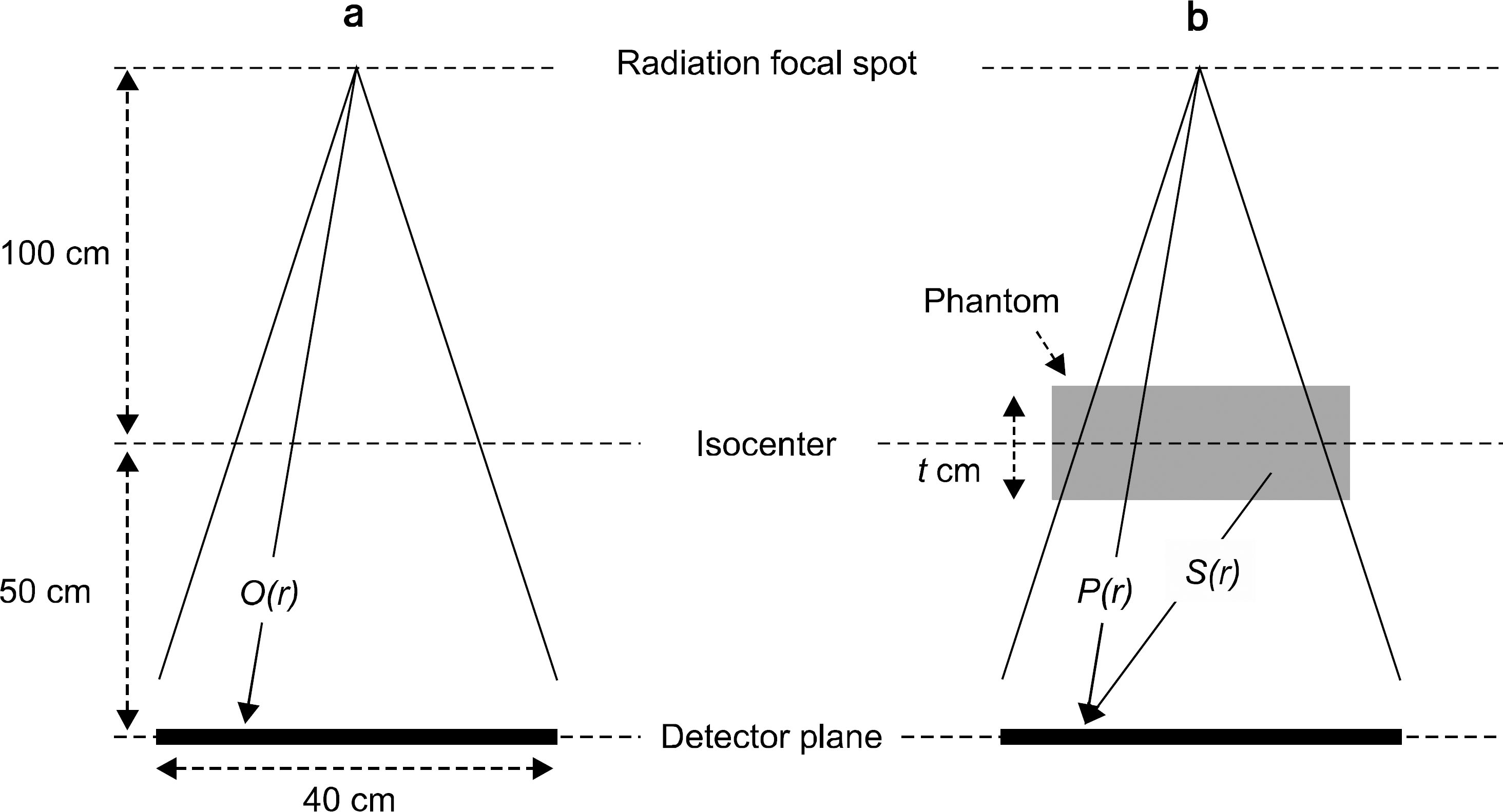
Fig. 3.
(a) The comparison of horizontal SPR profiles for different phantoms having four thicknesses, and (b) a SPR map measured using the phantom with the thickness of 20 cm.
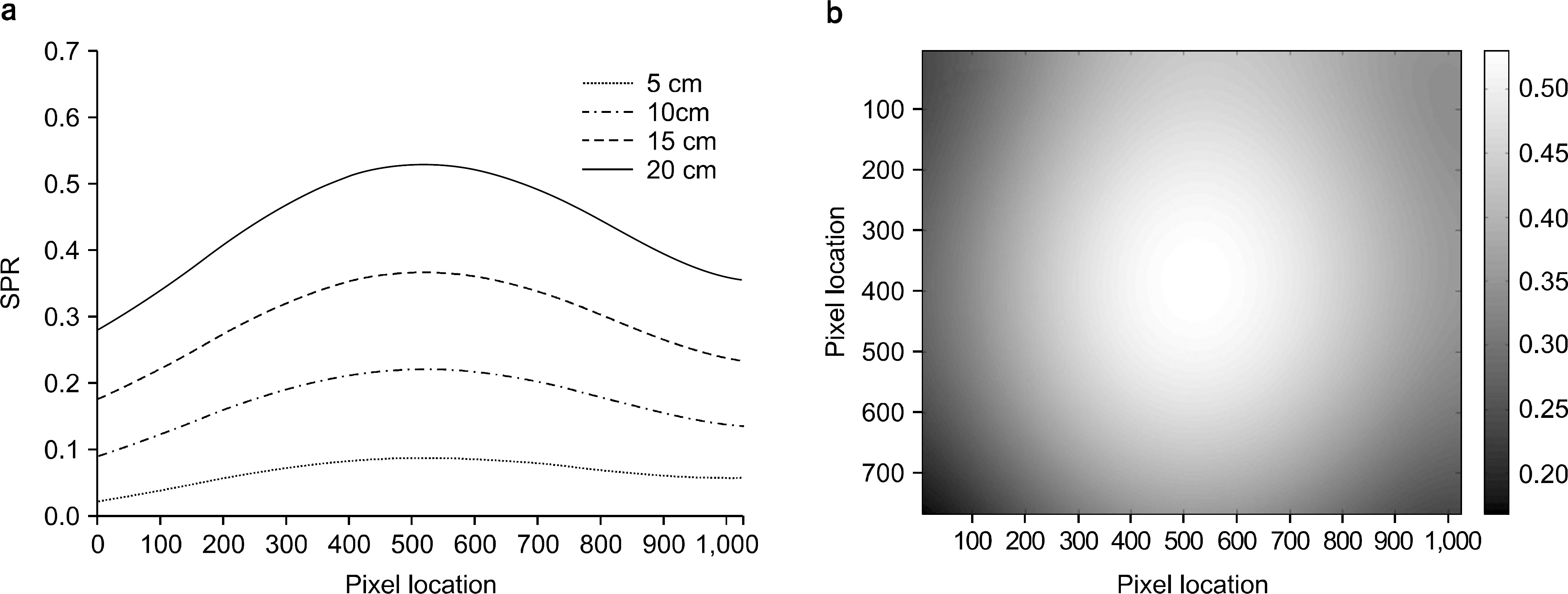
Fig. 4.
The line pair phantom images with (a) no correction and (b) SPR correction. The horizontal line profiles (see dashed lines in (a) and (b)) were compared in (c).

Fig. 5.
The comparison of (a) un-corrected and (b) corrected MV projection images of the pelvis. Five ROIs were marked on both images for the contrast evaluation.

Fig. 6.
The comparison of un-corrected and corrected histograms for (a) the line pair phantom and (b) the pelvis imag
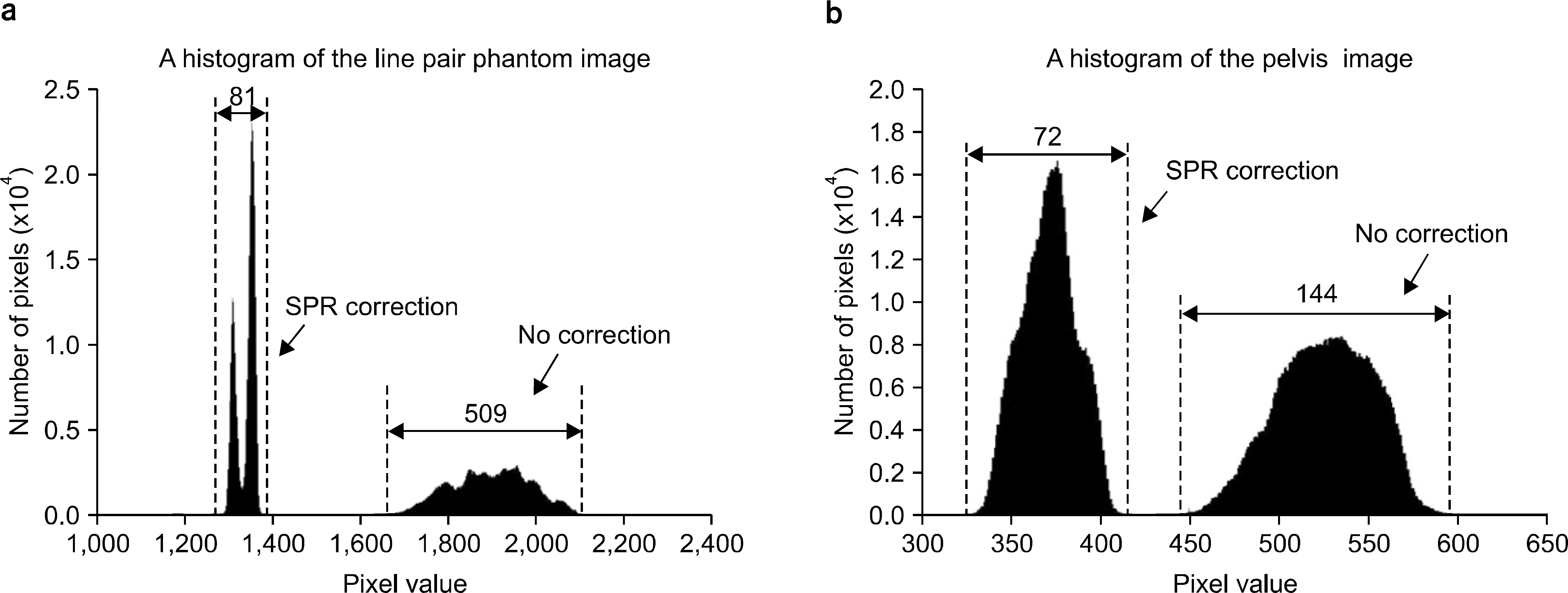
Table 1.
The un-attenuated ratio water phantoms with four diffe os of 6MV x-ray beams for solid-erent thicknesses (calculated).
| t (cm) | e−μeff⋅t |
|---|---|
| 5 | 0.7374 |
| 10 | 0.5499 |
| 15 | 0.4143 |
| 20 | 0.3151 |
Table 2.
The normalized contrast values of image patches of five ROIs (ΔC: contrast difference of no corrected and SPR corrected ROIs).
| CROI | ΔC | ||
|---|---|---|---|
| no correction | SPR correction | ||
| ROI1 | 0.111 | 0.250 | 0.139 |
| ROI2 | 0.167 | 0.194 | 0.027 |
| ROI3 | 0.132 | 0.139 | 0.007 |
| ROI4 | 0.118 | 0.181 | 0.063 |
| ROI5 | 0.139 | 0.222 | 0.083 |
| Mean | 0.133 | 0.197 | 0.064 |
 XML Download