Abstract
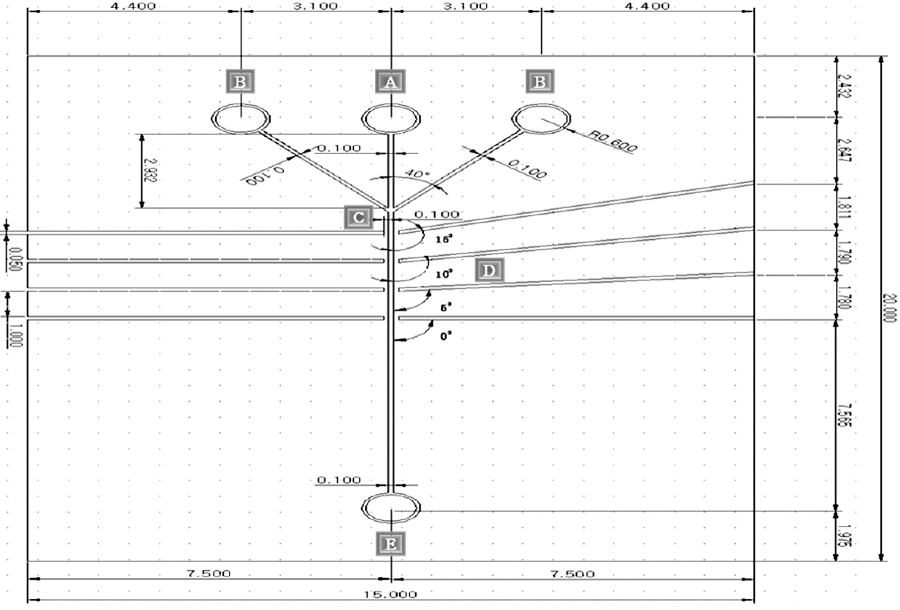
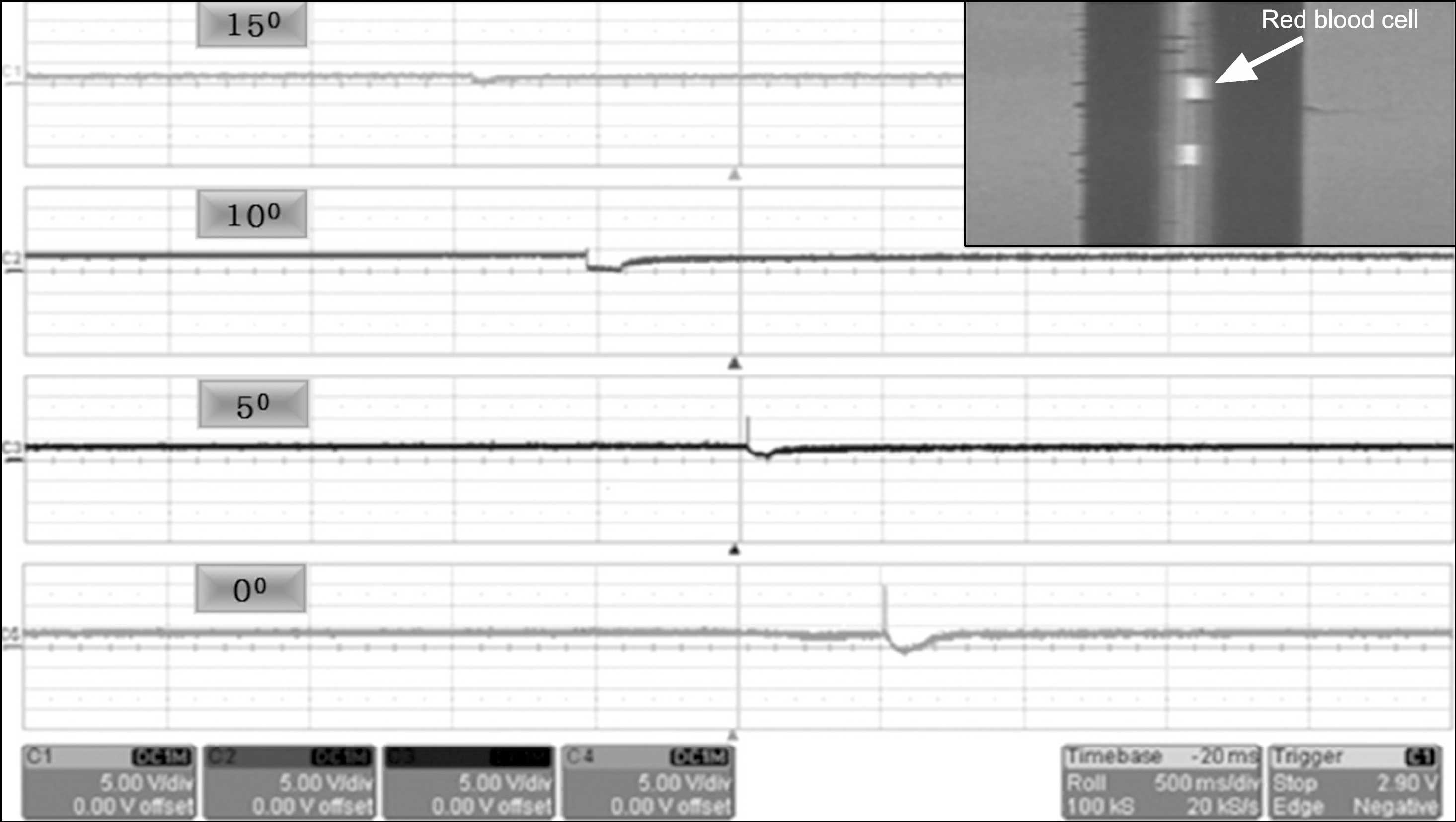
Next future, The bio technology will be a rapidly developing. This paper is the scattering beam measurement of the red blood cell (RBC) and the fabrication of the micro cell biochip using the bio micro electro mechanical system (Bio-MEMS) process technology. The Major process method of Bio-MEMS technology was used the buffered oxide etchant (BOE), electro chemical discharge (ECD) and ultraviolet sensitive adhesives (UVSA). All experiments were the 10 times according to the process conditions. The experiment and research are required the ultraviolet expose, the micro fluid current, the cell control and the measurement of the output voltage Vpp (peak to peak) waveform by scattering angles. The transmitting and receiving of the laser beam was used the single mode optical fiber. The principles of the optical properties are as follows. The red blood cells were injected into the micro channel. The single mode optical fiber was inserting in the guide channel. The He-Ne laser beam was focusing in the single mode optical fiber. The transmission He-Ne laser beam is irradiating to the red blood cells. The manufactured guide channel consists of the four inputs and the four outputs. The red blood cell was allowed with the cylinder pump. The output voltage Vpp waveform of the scattering beam was measured with a photo detector. The receiving angle of the output optical fiber is 0o, 5o, 10o, 15o. The magnitude of the output voltage Vpp waveform was measured in the decrease according to increase of the reception angles. The difference of the output voltage Vpp waveform is due differences of the light transmittance of the red blood cells.
Go to : 
References
1. The Korean Federation of Science and Technology Societies. 2020 year bio economic opening. Science and technology. 375(42–45):1599–7340. 2000.
2. Jing XM, Chen D, Fang DM, Huang C, Liu JQ. Multilayer microstructure fabrication by combining bulk silicon micromachining and UV-LIGA technology. Microelectronics J. 38:120–124. 2007.

3. Bu MI, T Melvin, GJ Ensell: A new masking technology for deep glass etching and its microfluidic application. Sensors and Actuators A. 115:476–482. 2004.
4. Hu CY, Lo SL, Li CM, Kuan WH. Treating chemical mechanical polishing (CMP) wastewater by electro coagulation flotation process with surfactant. J Hazardous Mater. 120:15–20. 2005.
5. 정안목, 전의식, 김철호: A study on the metal-glass bonding using ultra-sonic. J the Semiconductor & Display Technology. 10:(. (2):):. June. 2011.
6. Zbigniew D, Paul R, Harry C. Flow cytometry. Academic press (2nd): part A-B (. 1995.
7. David PS, Christopher TC, Stephen CJ, Michael R. Microchip flow cytometry using electro kinetic focusing. Analytical Chemistry. 71(19):4173–4177. 1999.
8. Kim S, Williams RT, Lee HK, Midori M. Turner size characterization of magnetic cell sorting micro beads using flow field flow fractionation and photon correlation spectroscopy. J Magnetism and Magnetic Materials. 194:248–253. 1999.
9. Daniel S, Young AM, Gray ML, Senturia SD. A micro fabricated flow chamber for optical measurements in fluids. IEEE: 219–224 (. 1993.
10. Yoo DS, Sim KS. Track distribution of recoil protons in PN-3 dosimeters etched in NaOH solution. Korean Society of Medical Physics. 2(2):129–139. 1991.
11. Lee BY, Yoon HG, Hyun KS, Kwon YH, Yun IG. Investigation of manufacturing variations of planar InP/InGaAs avalanche photodiodes for optical receivers. Microelectronics J. 35:635–640. 2004.

12. Al-Mufti R, Hambley H, Albaiges G, Lees C, Nicolaides KH. Increased fetal erythroblast in women who subsequently develop preeclampsia. Hum Reprod. 15:1624–1628. 2000.
13. 신동오, 홍성언, 이병용, 이명자: The construction of solid state detector system using commercially available diode and its application. Korean Society of Medical Physics. 1(1):91–95. 1990.
14. Jian J, Lili C, Chengrong S, Fu XZ. A study of the effect of PTFE electret on fibroblast cell cycle with flow cytometry (FCM). Proceedings of 10th International Symposium on Electrets: 171–173 (. 1999.
Go to : 


 PDF
PDF ePub
ePub Citation
 Print
Print


 XML Download