Abstract
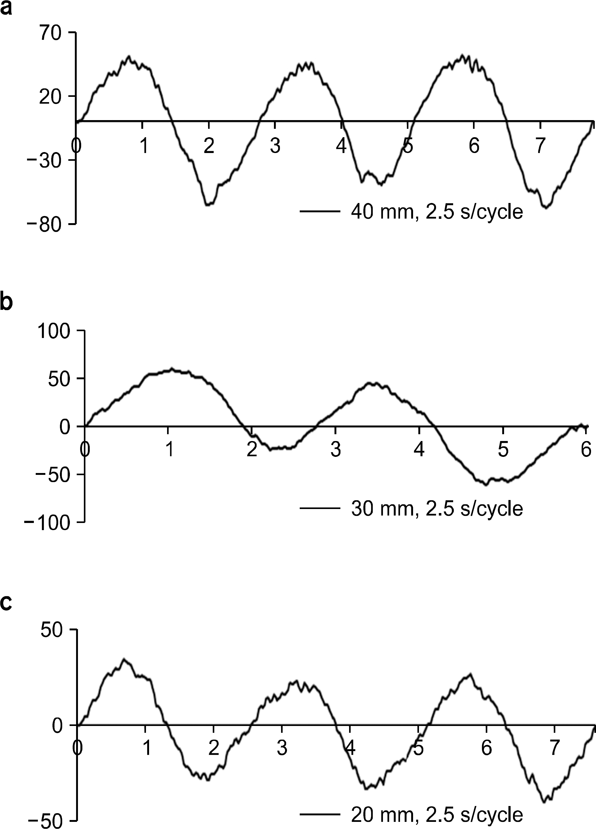
In this study, we developed and evaluated the patient respiration training method which can help to avoid the problems for the limitation of RGRT applicable patient cases. By using the MEMS (micro-electro-mechanical-system) acceleration sensor, we measured movement of motion phantom. We had compared the response of MEMS with commercially introduced real time patient monitoring (RPM) system. We measured the response of the MEMS with 1 dimensional motion phantom movement for 2.5, 3.0, 3.5 second of period and the 2.0, 3.0, 4.0 cm of the amplitudes. The measured period error of the MEMS system was 0.6∼6.0% compared with measured period using RPM system. We found that the shape of MEMS signals were similar with RPM system. From this study, we found the possibility of MEMS as patient training system.
References
1. Keal PJ, Mageras GS, Balter M, et al. The management of respiratory motion in radiation oncology report of AAPM Task Group. Med Phys. 33:3874–3881. 2006.
2. Britton KR, Starkschall G, Tucker SL, et al. Assessment of gross tumor volume regression and motion changes during radiotherapy for non-small-cell lung cancer as measured by four-dimensional computed tomography. Int J Radiat Oncol Biol Phys. 68:1036–1046. 2007.

3. Chang J, Mageras GS, Yorke E, et al. Observation of interfractional variations in lung tumor position using respiratory gated and ungated megavoltage cone-beam computed tomography. Int J Radiat Oncol Biol Phys. 67:1548–1558. 2007.

4. Weiss E, Wijesooriya K, Dill SV, et al. Tumor and normal tissue motion in the thorax during respiration: Analysis of volumetric and positional variations using 4D CT. Int J Radiat Oncol Biol Phys. 67(1):296–307. 2007.

5. Rietzel E, Liu AK, Chen GTY, et al. Maximum-intensity volumes for fast contouring of lung tumors including respiratory motion in 4DCT planning. Int J Radiat Oncol Biol Phys. 71(4):1245–1252. 2008.

6. Ju SG, Hong C, Huh W, et al. Development of an offline based internal organ motion verification system during treatment using sequential cine EPID images. Korean J Med Phys. 23(2):91–98. 2012.
7. Webb S. Motion effects in (intensity modulated) radiation therapy: a review. Phys Med Biol. 51(13):R403–R425. 2006.

8. Minohara S, Kanai T, Endo M, et al. Respiratory gated irradiation system for heavy-ion radiotherapy. Int J Radiat Oncol Biol Phys. 47(4):1097. 2000.

9. Liu HH, Koch N, Starkschall G, et al. Evaluation of internal lung motion for respiratory-gated radiotherapy using MRI: Part II – Margin reduction of internal target volume. Int J Radiat Oncol Biol Phys. 60:1473–1483. 2004.
10. Mah D, Hanley J, Rosenzweig KE, et al. Technical aspects of the deep inspiration breath-hold technique in the treatment of thoracic cancer. Int J Radiat Oncol Biol Phys. 48(4):1175–1185. 2000.

11. Berson AM, Emery R, Rodriguez L, et al. Clinical experience using respiratory gated radiation therapy: Comparison of free-breathing and breath-hold techniques. Int J Radiat Oncol Biol Phys. 60(2):419–426. 2004.

13. Shirato H, Harada T, Harabayashi T, et al. Feasibility of insertion/implantation of 2.0-mm-diameter gold internal fiducial markers for precise setup and real-time tumor tracking in radiotherapy. Int J Radiat Oncol Biol Phys. 56(1):240–247. 2003.

15. George R, Chung TD, Vedam SS, et al. Audiovisual biofeedback for respiratory-gated radiotherapy: impact of audio instruction and audio-visual biofeedback on respiratory-gated radiotherapy. Int J Radiat Oncol Biol Phys. 65(3):924–933. 2006.

16. Neicu T, Berbeco R, Wolfgang J, et al. Synchronized moving aperture radiation therapy (SMART): improvement of breathing pattern reproducibility using respiratory coaching. Phys Med Biol. 51(3):617–636. 2006.

17. Jeong JD. Micro Electro Mechanical System, MEMS. In company: (. 2011. ). p. 78–83.
18. Zeng H, Zhao Y. Sensing movement: microsensors for body motion measurement. Sensors. 11(1):638–660. 2011.

19. http://www.standardimaging.com/,STANDARDIMAGINEphantomproducthome'respiratorygatingplatform'
Fig. 1.
MEMS acceleration sensor communication architecture (UM1017 User manual, STEVAL-MKI062V2 communication protocol).

Fig. 2.
Real MEMS signal displayed in the computer monitor (amplitude: 40 mm, velocity: 2.0 s/cycle).
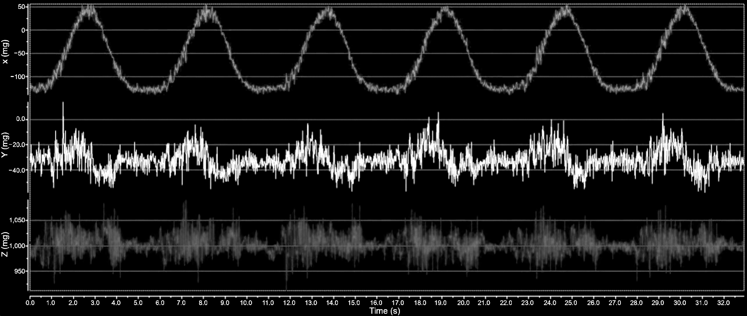
Fig. 3.
(a) Gravity acquired using the MEMS acceleration sensor, (b) velocity reconstructed by using equation (1), (c) position reconstructed by using equation (2) (amplitude: 30 mm, velocity: 3.5 s/cycle).
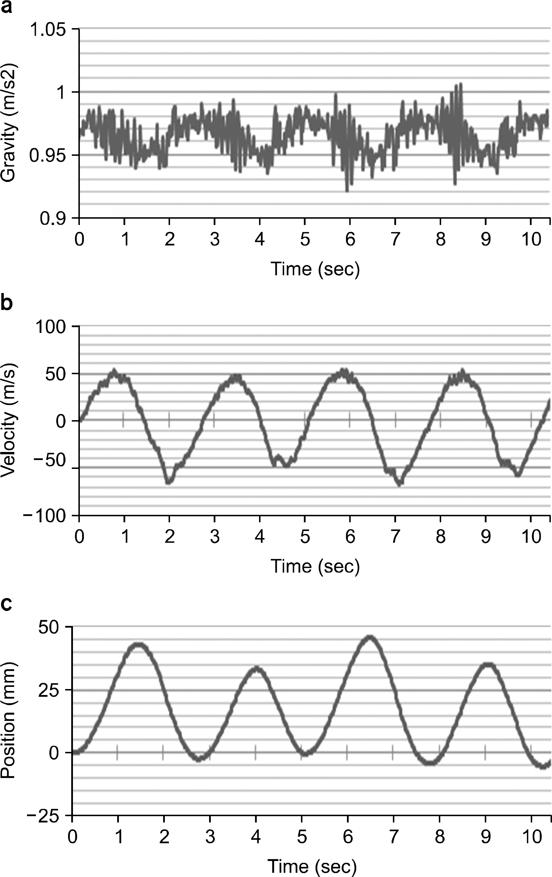
Fig. 4.
The motion of the respiratory Gating Platform (RGP) was measured by using the RPM system, independently.
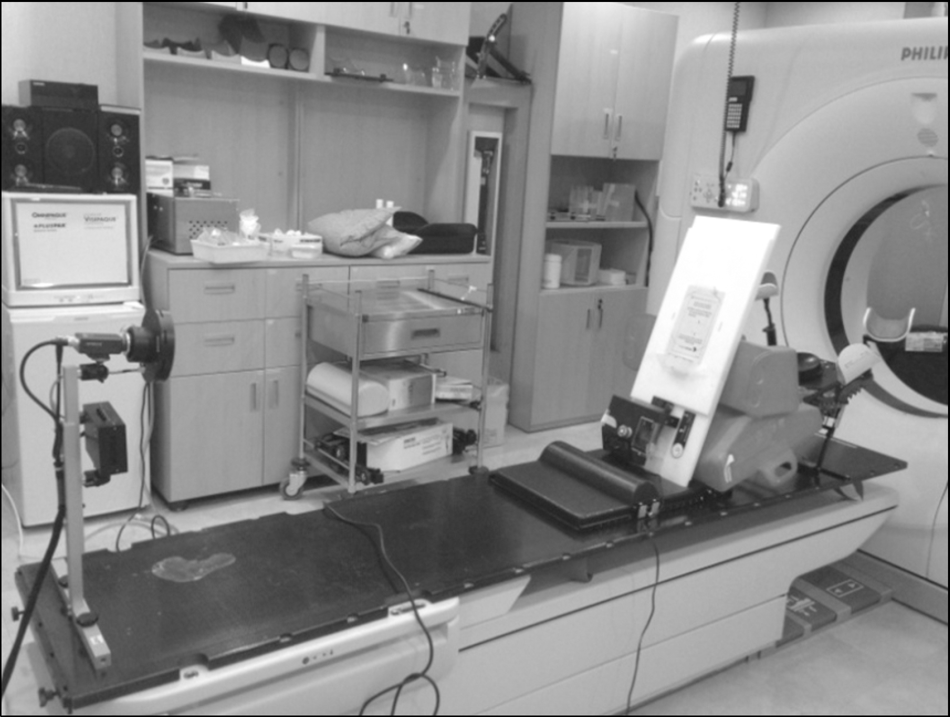
Fig. 6.
MEMS signal compared with RPM system. (a) 40 mm, 2.5 s/cycle MEMS signal (b) 40 mm, 2.5 s/cycle RPM system signal (c) 20 mm, 3.0 s/cycle MEMS signal (d) 20 mm, 3.0 s/cycle RPM system signal (e) 40 mm, 3.5 s/cycle MEMS signal (f) 40 mm, 3.5 s/cycle RPM system signal.
Table meas
e 1. Average sured with MEM cycle of 1D MS accelerati motion phanto ion sensor and om which was d their errors.


 PDF
PDF ePub
ePub Citation
 Print
Print


 XML Download