

 PDF
PDF ePub
ePub Citation
 Print
Print


Abstract
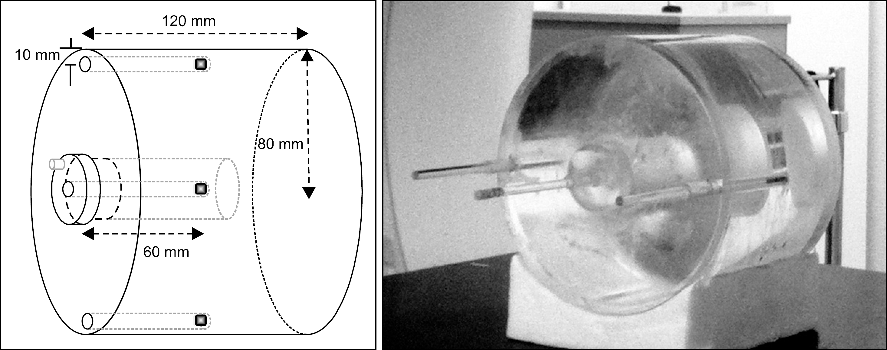
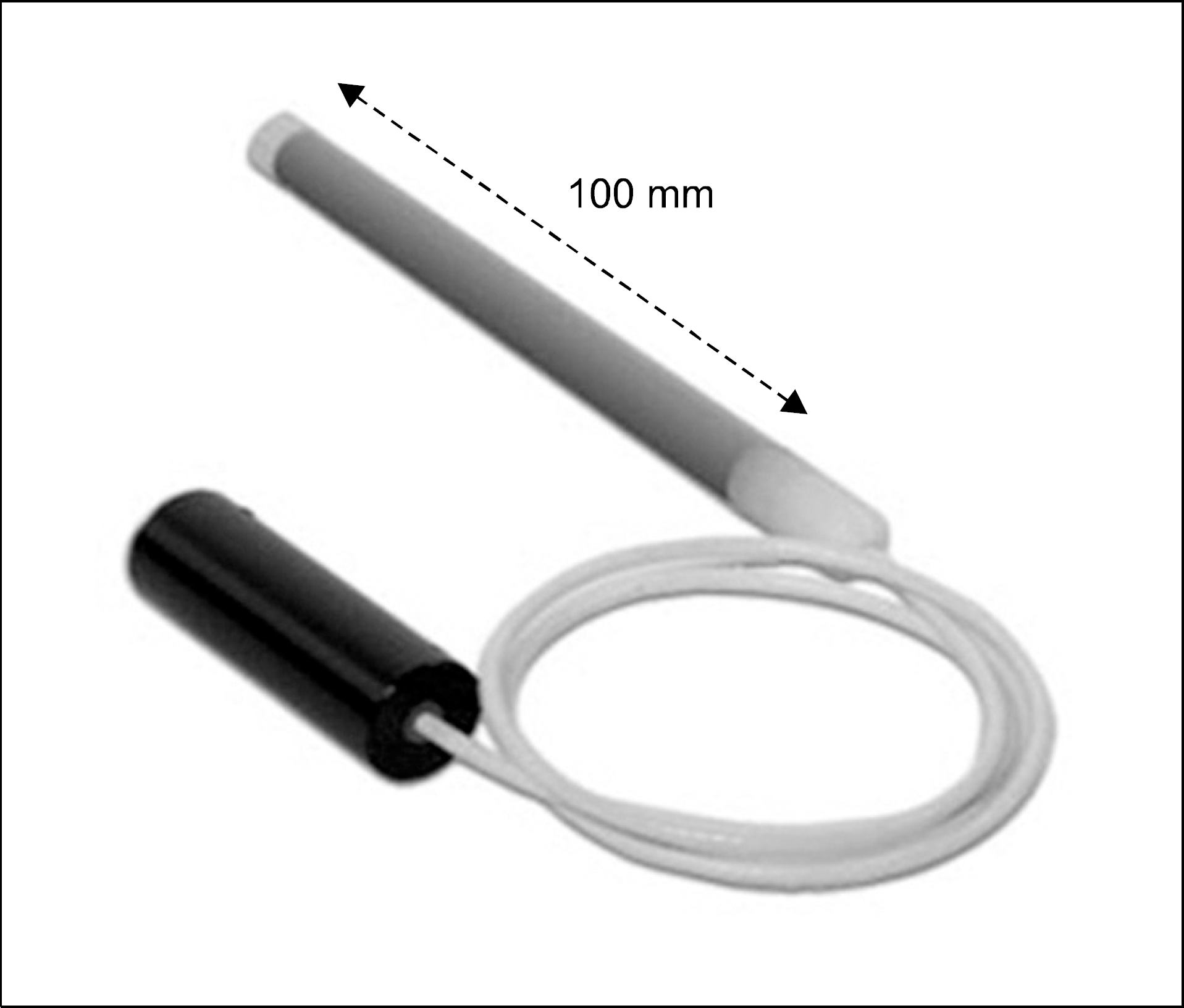
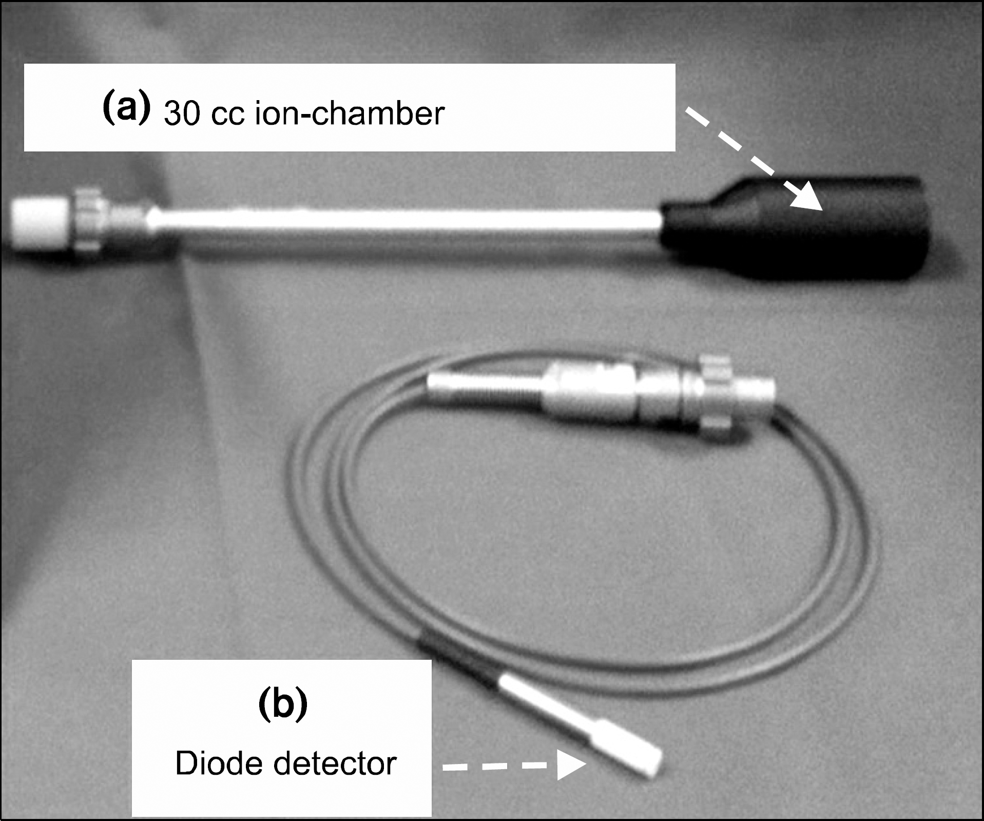
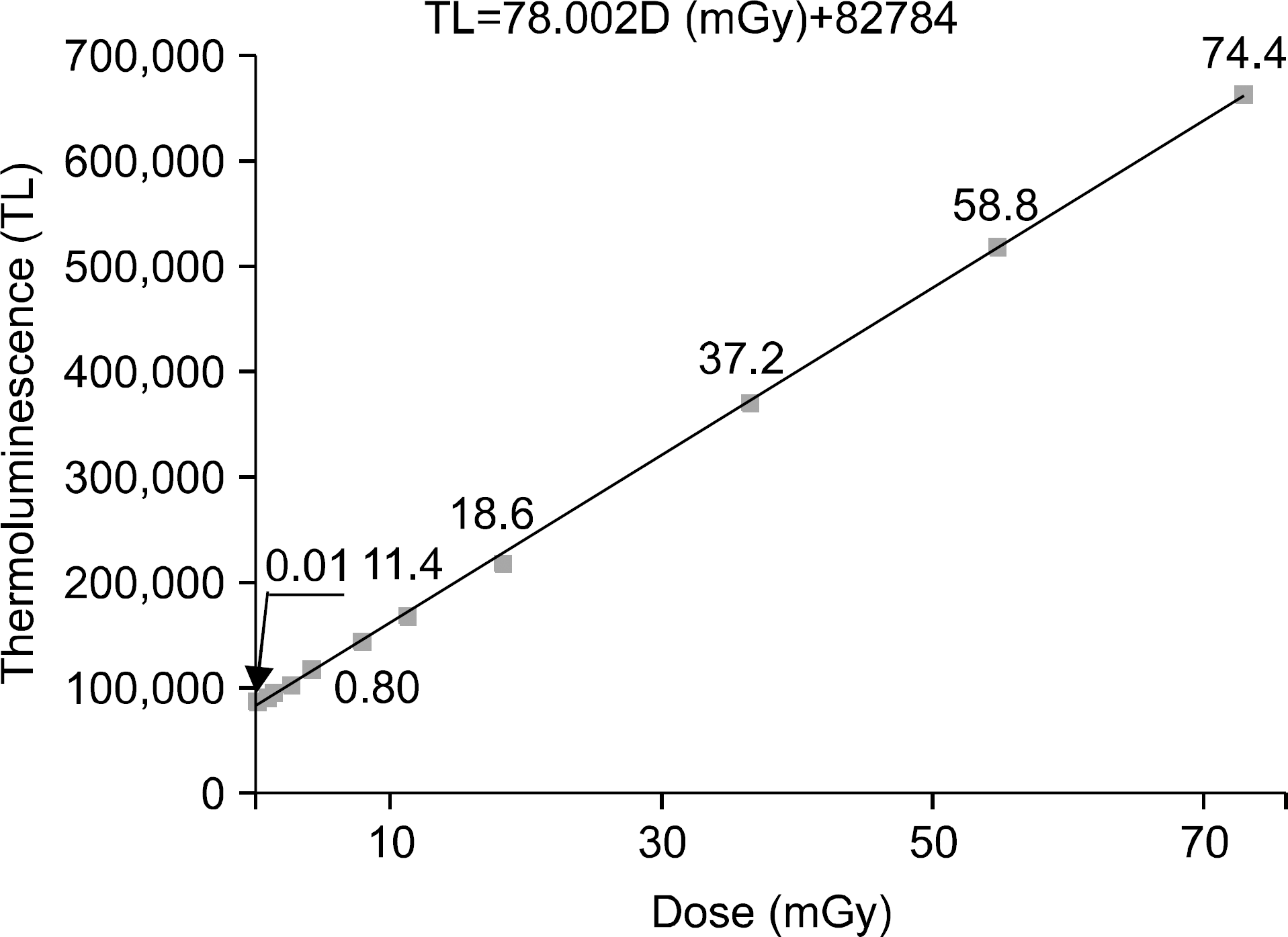
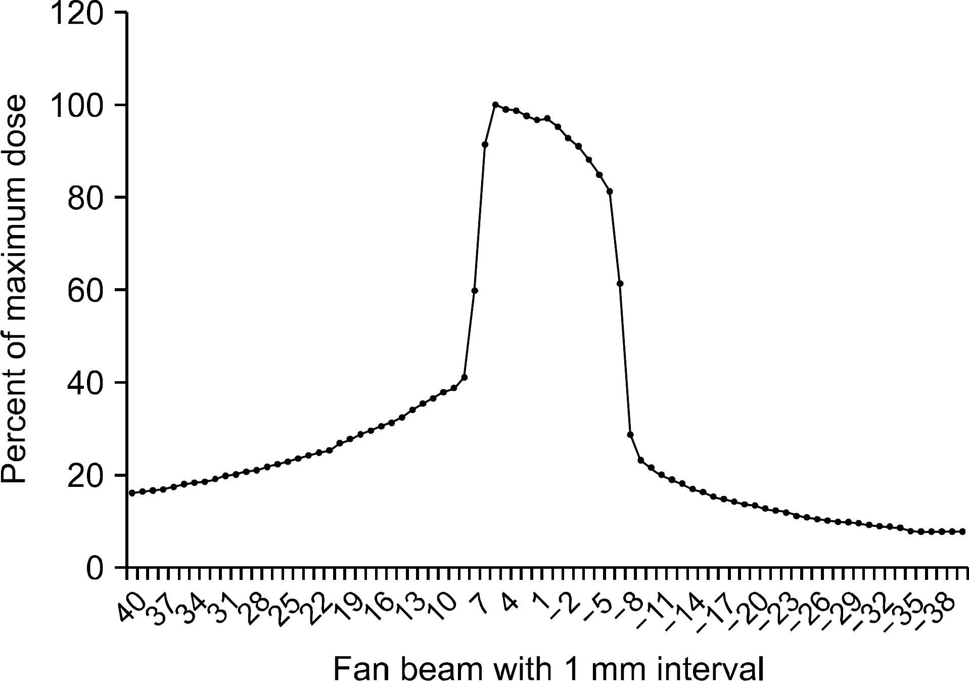
Recently, the uses of Multi-Detector Computed Tomography (MDCT) for radiation treatment simulation and planning which is used for intensity modulated radiation therapy with high technique are increasing. Because of the increasing uses of MDCT, additional doses are also increasing. The objective of this study is to evaluate the absorbed dose of body and skin undergoing in MDCT scans. In this study, the exposed dose at the surface and the center of the cylindrical water phantom was measured using an pencil ionization chamber, 30 cc ionization chamber and TL Powder. The results of MDCT were 31.84 mGy, 33.58 mGy and 32.73 mGy respectively. The absorbed dose at the surface showed that the TL reading value was 33.92 mGy from MDCT. These results showed that the surface dose was about 3.5% from the MDCT exposure higher than a dose which is located at the center of the phantom. These results mean that the total exposed dose undergoing MDCT 4 times (diagnostic, radiation therapy planning, follow-up et al.), is about 14 cGy, and have to be considered significantly to reduce the exposed dose from CT scan.
References
1. Hu H, He HD, Foley WD, Fox SH. Four Multidetector-row helical CT: image quality a nd volume coverage spee d. Radiology. 215:55–62. 2000.
2. Dawson P. Patient dose in multislice CT: Why is it increasing and doe s it ma tter? Br J Radiol 77: S 10-S 13 (. 2004.
3. Yates SJ, Pike LC, Goldstone KE. Effect of multislice scanners on patient dose from routine CT examinations in East Anglia. Br J Ra diol. 77:472–478. 2004.

4. Kwon SO, Dong KR, Kweon DC, et al. Estimate of Radiation Doses in MDCT Using Patient Weight. Korean J Med Phys. 21(3):246–252. 2010.
5. 김상연, 한진우, 박인우. Cone Beam CT와 일반 CT의 흡수선량 및 유효선량 비교평가.대한구강안면방사선학회지. 38:7–15. 2008.
6. Seoung YH, Kim YO, Choe BY. Re duc ing of cr aniofac ial radiation dose using automatic exposure control t e ch n ique in the 64 multi-de tector computed tomography. Kor ean J Med Phys. 21(2):137–144. 2010.
7. Lee CL, Kim HJ, Jeon SS, et al. Comp arison radiation dose in the mea sur ement of mdc t radiation dose ac cor ding to correction of temperatures and pressure, and calibration of ionization chamb er. Kor ean J Med Phys. 19(1):49–55. 2008.
8. 김문찬, 임종석, 박형로, 김유현:컴퓨터 단층 촬영시 환자 피폭 선량에 관한 연구.방사선 기술과학. 27:21–27. 2007.
9. 윤재혁, 이광원, 조영기 등:두경부(Head & Neck) CT검사 시 장기의 유효선량 측정.방사선기술과학. 34(2):105–116. 2011.
10. Scaf G, Lurie AG, Moisier KM, et al. Dosime try a nd cost of imaging osse ointe gr ated implants with film-based and computed tomography. Oral Surg Oral Med Oral Pathol Oral Radiol Endod. 83:41–48. 1997.
11. Task Group No.66: Quality assuar ance for computed-tomography simulators and th e computed-tomog raph y-simul ation process, AA PM Radiation Thera py C ommittee, US A. 2003.
12. Zhou H, Boone JM. Monte Ca rlo eva luation of CTDI∞ in infinitely long cylinders of water, polysthylene and PMMA with diameters from 10 mm to 500 mm. Med Phys. 35:2424–2431. 2008.
13. 최태진, 이호준, 예지원 등. LiF:Mg, Cu, P열형광선량계의 선량특 성을 이용한 눈가림법에 의한 출력선량 평가.의학물리. 20(4):308–316. 2009.
 XML Download