

 PDF
PDF ePub
ePub Citation
 Print
Print


Abstract
Correct target volume delineation is an important part of radiosurgery treatment planning process. We designed head phantom and performed target delineation to evaluate the volume differences due to radiosurgery treatment planning systems and image acquisition system, CT/MR. Delineated mean target volume from CT scan images was 2.23±0.08 cm3 on BrainSCAN (NOVALS), 2.13±0.07 cm3 on Leksell gamma plan (Gamma Knife) and 2.24±0.10 cm3 on Multi plan (Cyber Knife). For MR images, 2.08±0.06 cm3 on BrainSCAN, 1.94±0.05 cm3 on Leksell gamma plan and 2.15±0.06 cm3 on Multi plan. As a result, Differences of delineated mean target volume due to radiotherapy planning system was 3% to 6%. And overall mean target volume from CT scan images was 6.36% larger than those of MR scan images.
REFERENCES
1. Klein EE, Hanley J, Bayouth J, et al. Task Group 142 report: Quality assurance of medical accelerators. Med Phys. 36(9):4197–4212. 2009.
2. Kutcher GJ, Coia L, Gillin M, Hanson WF, et al. Comprehensive QA for radiation oncology: Report of AAPM Radiation Therapy Committee Task Group 40. Med Phys. 21(4):581–618. 1994.

3. Robar JL, Clark BG. The use of radiographic film for linear accelerator stereotactic radiosurgical dosimetry. Med Phys. 26(10):2144–2150. 1999.
4. 이경남, 이동준, 서태석. 자기공명영상기반 겔 선량측정법을 이용한 3차원적 목표 중심점 점검기술. 의학물리. 22(1):35–41. 2011.
5. Mazzara GP, Velthuizen RP, Pearlman JL, et al. Brain tumor target volume determination for radiation treatment planning through automated MRI segmentation. Int J Radia Oncol Biol Phys. 59(1):300–312. 2004.
6. Rasch C, Keus R, Pameijer FA, et al. The potential impact of CT-MRI matching on tumor volume delineation in advanced head and neck cancer. Int J Radia Oncol Biol Phys. 39(4):841–848. 1997.
7. Caldwell CB, Mah K, Ung YC, et al. Observer variation in contouring gross tumor volume in patients with poorly defined Non-small-cell lung tumors on CT: The impact of 18FDG-hybrid pet fusion. Int J Radia Oncol Biol Phys. 51(4):923–931. 2001.
8. Kouwenhoven E, Giezen M, Struikmans H. Measuring the similarity of target volume delineations independent of the number of observers. Phys Med Bio. 54(9):2863–2873. 2009.
9. Weltens C, Menten J, Feron M, et al. Interobserver variation in gross tumor volume delineation of brain tumor on computed tomography and impact of magnetic resonance imaging. Radiotherapy & Oncology. 60(1):49–59. 2001.
10. Breen SL, Publicover J, De Silva S, et al. Intraobserver and Interobserver variability in GTV Delineation on FDG-PETCT images of Head and Neck Cancers. Int J RadiaOncol- BiolPhys. 68(3):763–770. 2007.
11. Geets X, Daisne JF, Tomsej M, et al. Impact of the type of imaging modality on target volumes delineation and dose distribution in Pharyngo-laryngeal squamous cell carcinoma: comparison between pre- and per treatment studies. Radiotherapy & Oncology. 78(3):291–297. 2006.
12. Ackerly T, Andrews J, Ball D, et al. Discrepancies in volume calculations between different radiotherapy treatment planning systems: Australas Phys Eng Sci Med. 26(2):91–93. 2003.
Fig. 1.
Small target volume location in custom designed head phantom. (a) Axial view. (b) Oblique view.
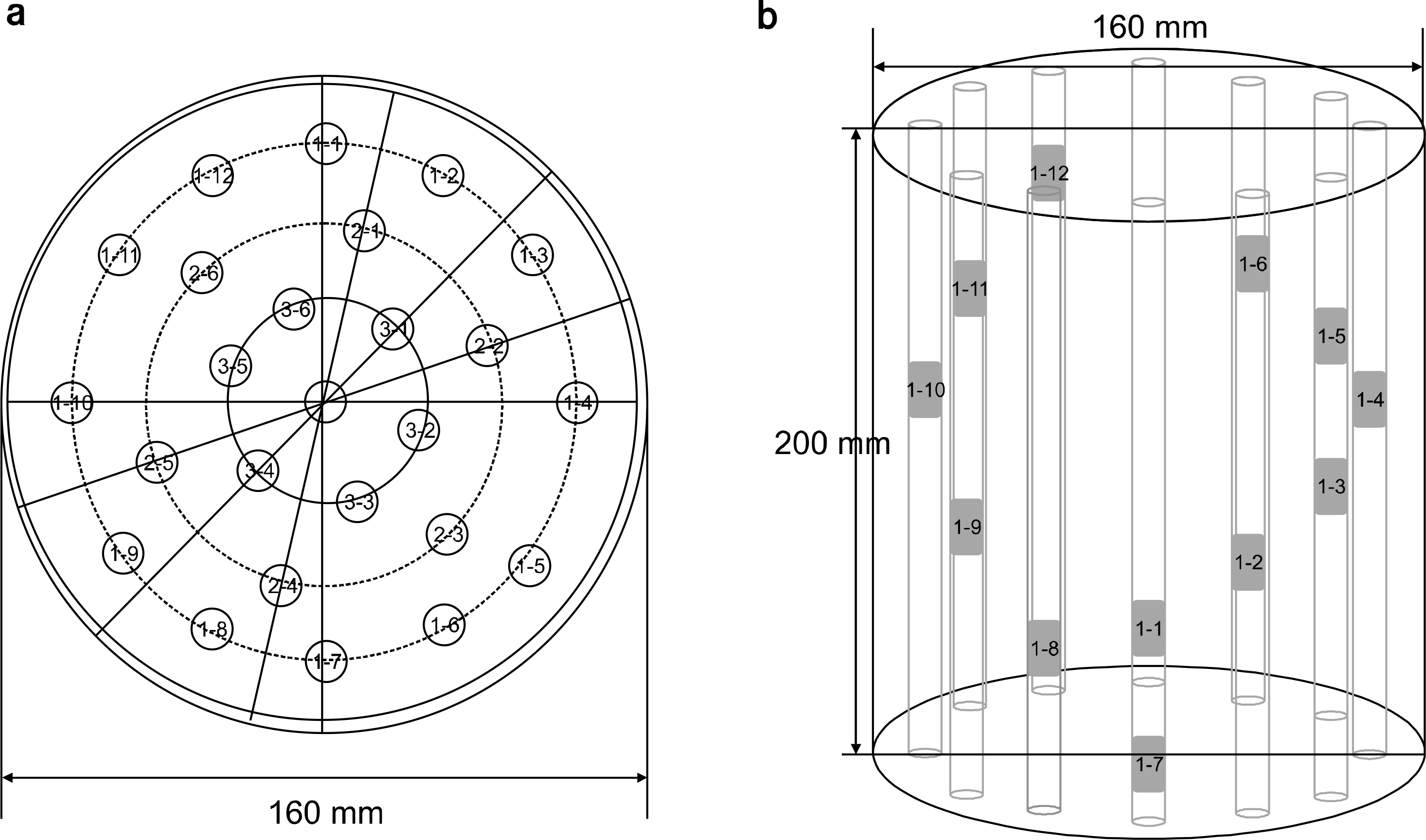
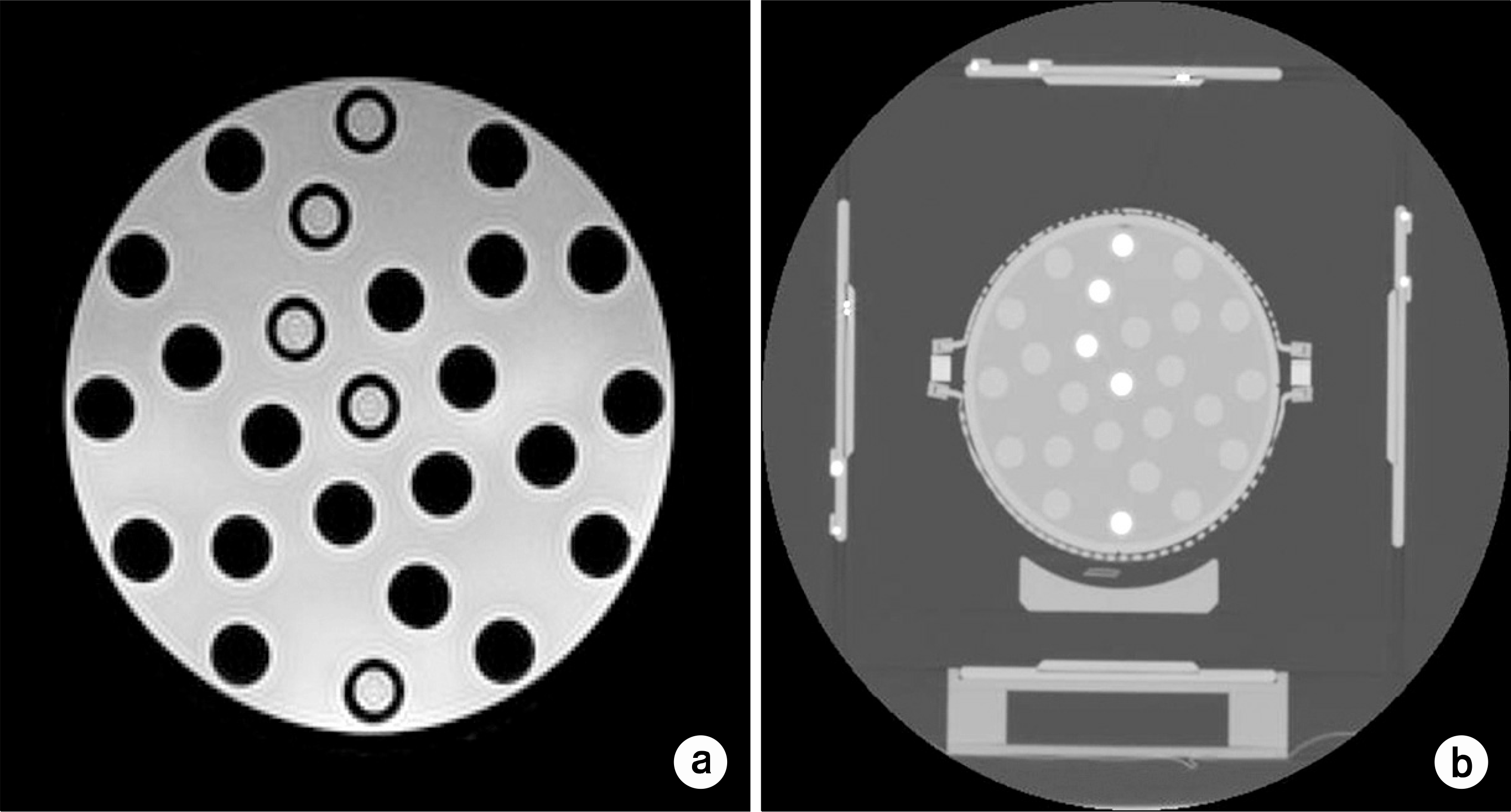
Fig. 3.
Comparison of small target volume acquired from (a) CT image (b) MRI Image depends on radiotherapy planning system.
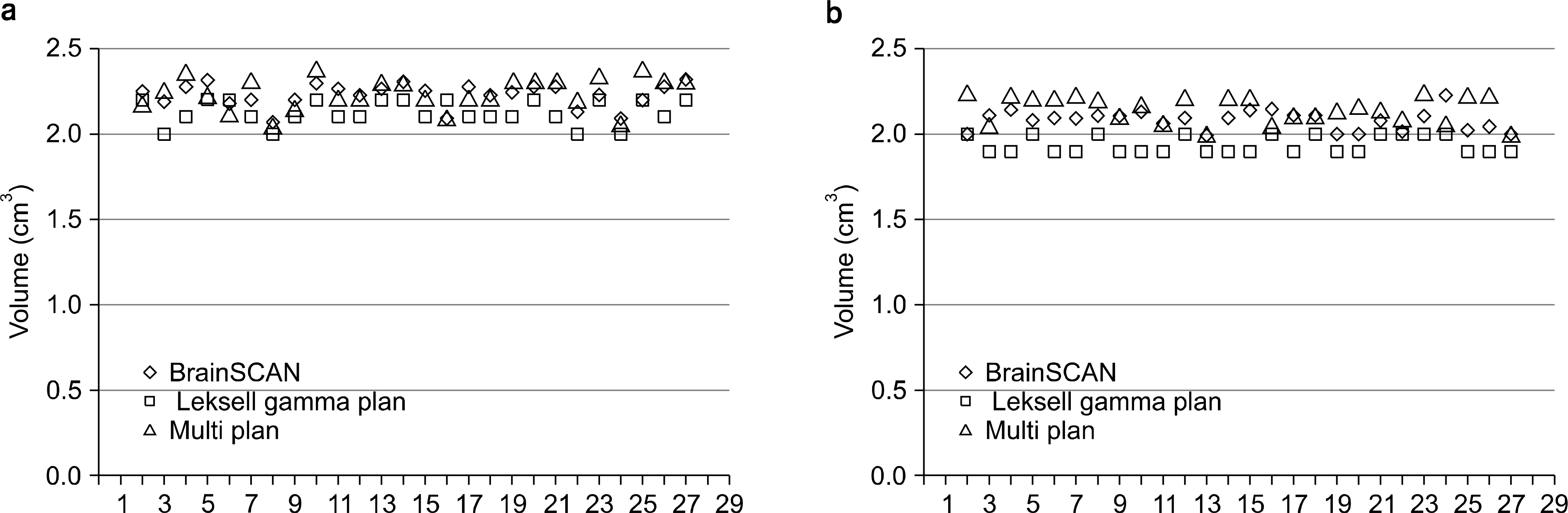
Fig. 4.
Comparison of small target volume in radiotherapy planning system depends on image acquisition system (CT1, MRI1: BrainSCAN, CT2, MRI2: Leksell gamma plan, CT3, MRI3: Multi plan).
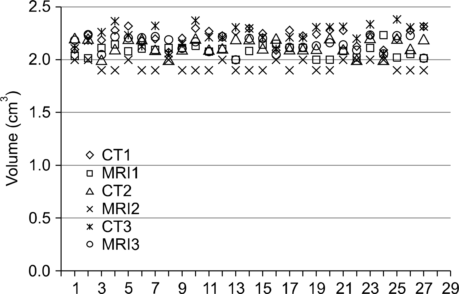
Table 1.
CT/MR image acquisition protocol for evaluate the delineated small target volume.
| CT | MRI | |
|---|---|---|
| Slice thickness | 2.0 mm | 2.0 mm |
| Inter slice gap | Not gap | No gap |
| Field of View (FOV) | 379 mm×379 mm | 250 mm×250 mm |
| Matrix size | 512×512 | 512×512 |
| TR/TE | 5,631 ms/105 ms | |
| Coil | Head coil used |
Table 2.
Delineated mean target volume from CT/MR scan images in radiosurgery planning system. (Mean±S.D) cm3
Table 3.
Target volume differences between CT/MR delineation depend on radiotherapy planning system.
Table 4.
Target volume differences depend on acquired CT/MRI.
 XML Download