

 PDF
PDF ePub
ePub Citation
 Print
Print


Abstract
The DICOM converter program of the Geant4 Monte Carlo simulation code for the application of radiotherapy was developed. We analysis the header part of the DICOM file and find various parameters, such as matrix size, pixel size, stored data bits, high bit, and padding values. Especially we evaluate every pixel value of the DICOM files. To conform the exact convert of the pixel values, we developed the verify program. As a result, the DICOM formats generated from difference CT vendors can be converted and verified for Genat4 calculations.
REFERENCES
1. Joh YG, Kim HD, Kim BY, et al. Calculation of energy spectra for electron beam of medical linear accelerator using GEANT4. Korean Journal of Medical Physics. 22(2):85–91. 2011.
2. Park SH, Jung WG, Rah JE, Park S, Suh TS. Evaluation of the secondary particle effect in inhomogeneous media for proton therapy using Geant4 based MC simulation. Journal of Medical Physics. 21(4):311–322. 2010.
3. Kimura A, Tanaka S, Aso T, et al. DICOM interface and visulization tool for Geant4-based dose calculation. Nuclear Science Symposium Conference Record 2005 IEEE Volume. 2:981–984. 2005.
4. Agostinelli S, Allison J, Amako K, et al. Geant4 developments and applications. IEEE Transactions on Nuclear Science. 53(1):270–278. 2006.
5. Kim BY, Kim HD, Kim SJ, Oh SA, Kang JK, Kim SK. Study on the 6MV photon beam characteristics and analysis method from medical linear accelerators using Geant4 medical Linac2 example. Journal of Medical Physics. 22(2):79–84. 2011.
6. GEANT4 Collaboration Version Geant4 9.6.0. Geant4 user's guide for application developers. CERN. 2013.
7. ICRU Report 46. Photon, Electron, Proton and Neutron Interaction data for Body Tissues. International Commission on Radiation Units and Measurement, bethesda, MD. 1992.
8. Agostinelli S, Allison J, Amako K, et al. Geant4 – a simulation toolkit. Nuclear Instruments and Methods A. 506(3):250–303. 2003.
Fig. 3.
Difference of bits stored. (a) bits stored=12 and High bits= 11 (b) bits stored=16 and high bit=15.
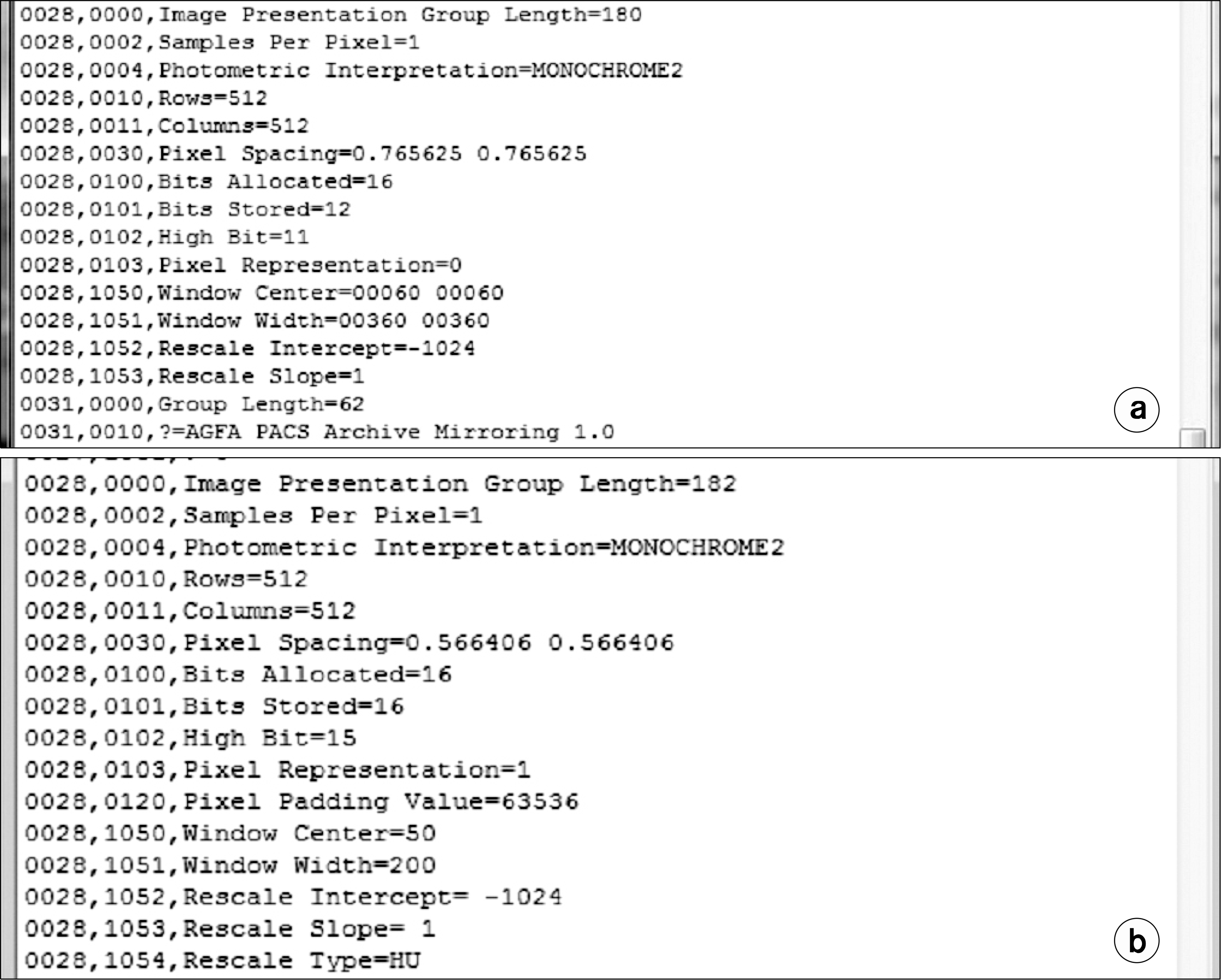
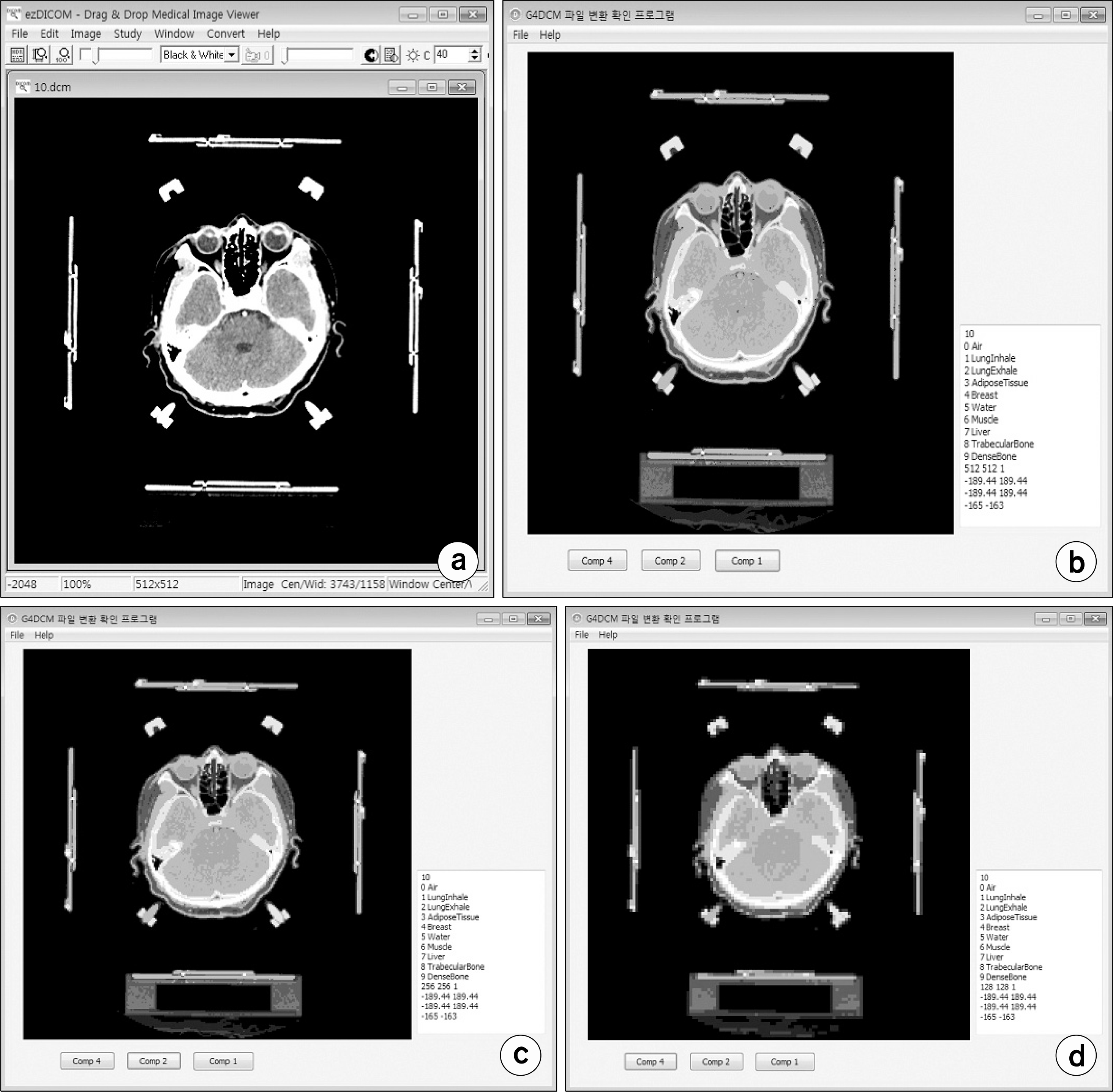
Fig. 4.
Evaluation of converted meta files. (a) original DICOM file (b) compressed file (compression=1) (c) compressed file (compression=2) (d) compressed file (compression=4).
 XML Download