

 PDF
PDF ePub
ePub Citation
 Print
Print


Abstract
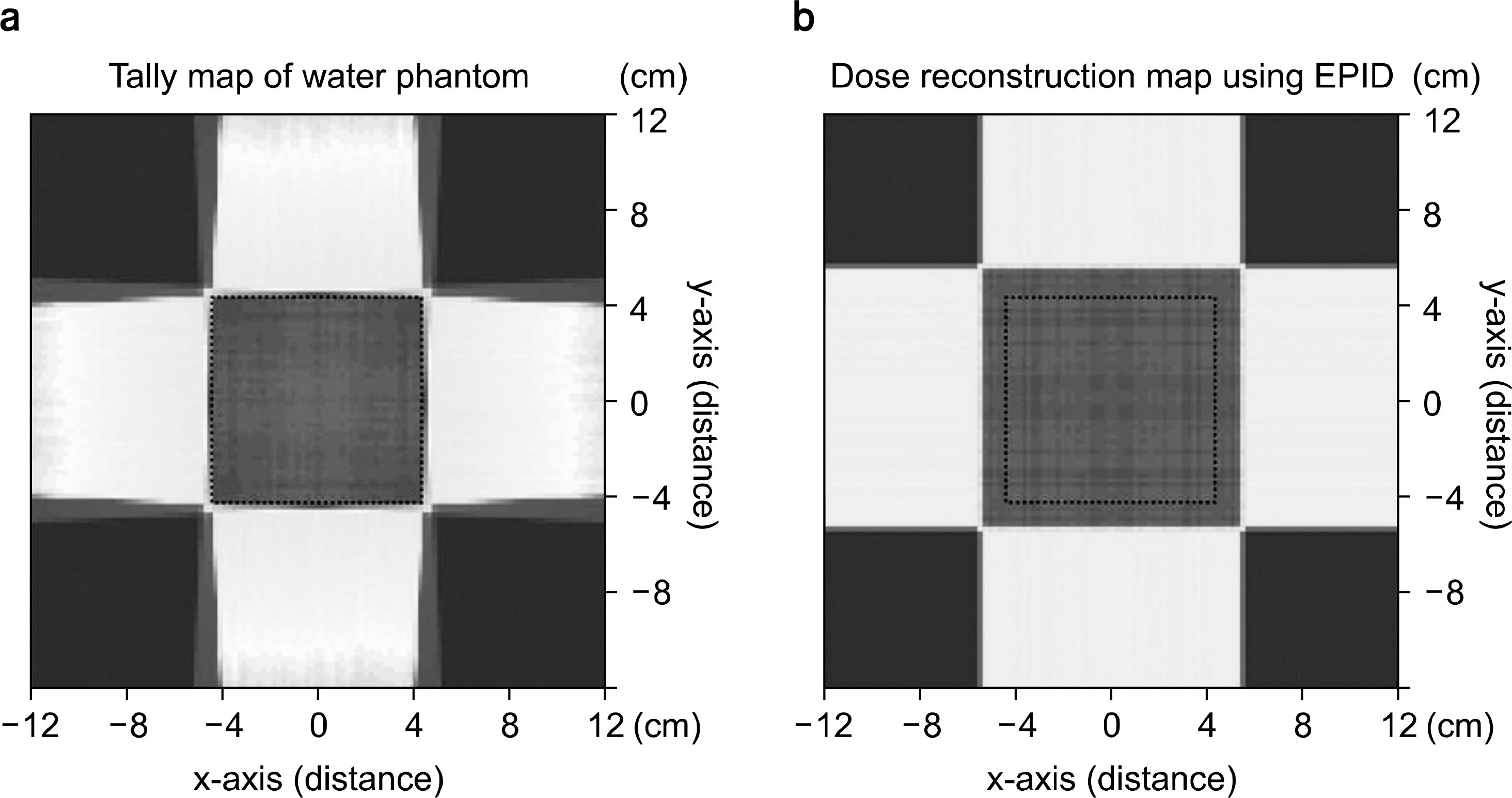
The purpose of this study is to evaluate and analyze the relationship between the external radiation dose reconstruction which is transmitted from the patient who receives radiation treatment through electronic portal imaging device (EPID) and the internal dose derived from the Monte Carlo simulation. As a comparative analysis of the two cases, it is performed to provide a basic indicator for similar studies. The geometric information of the experiment and that of the radiation source were entered into Monte Carlo n-particle (MCNPX) which is the computer simulation tool and to derive the EPID images, a tally card in MCNPX was used for visualizing and the imaging of the dose information. We set to source to surface distance (SSD) 100 cm for internal measurement and EPID. And the water phantom was set to be 100 cm of the source to surface distance (SSD) for the internal measurement and EPID was set to 90 cm of SSD which is 10 cm below. The internal dose was collected from the water phantom by using mesh tally function in MCNPX, accumulated dose data was acquired by four-portal beam exposures. At the same time, after getting the dose which had been passed through water phantom, dose reconstruction was performed using back-projection method. In order to analyze about two cases, we compared the penetrated dose by calibration of itself with the absorbed one. We also evaluated the reconstructed dose using EPID and partially accumulated (overlapped) dose in water phantom by four-portal beam exposures. The sum dose data of two cases were calculated as each 3.4580 MeV/g (absorbed dose in water) and 3.4354 MeV/g (EPID reconstruction). The result of sum dose match from two cases shows good agreement with 0.6536% dose error.
REFERENCES
1. Chang J, Obcemea CH, Sillanpaa J, Mechalakos J, Burman C. Use of EPID for leaf position accuracy QA of dynamic multi-leaf collimator (DMLC) treatment. Med Phys. 31(7):2091–2096. 2004.

2. Parent L, Seco J, Evans PM, Dance DR, Fielding A. Evaluation of two methods of predicting MLC leaf positions using EPID measurements. Med Phys. 33(9):3174–3182. 2006.
3. Vial P, Greer PB, Hunt P, Oliver L, Baldock C. The impact of MLC transmitted radiation on EPID dosimetry for dynamic MLC beams. Med Phys. 35(4):1267–1277. 2008.
4. Greer PB, Cadman P, Lee C, Bzdusek K. An energy fluence- convolution model for amorphous silicon EPID dose prediction. Med Phys. 36(2):547–555. 2009.
5. McDermott LN, Wendling M, van Asselen B, et al. Clinical experience with EPID dosimetry for prostate IMRT pretreatment dose verification. Med Phys. 33(10):3921–3930. 2006.
6. Spezi E, Lewis DG. Full forward Monte Carlo calculation of portal dose from MLC collimated treatment beams. Phys MedBiol. 47:377–390. 2002.
7. Paganetti H, Jiang H, Lee SY, Kooy HM. Accurate Monte Carlo simulations for nozzle design, commissioning and quality assurance for a proton radiation therapy facility. Med Phys. 31(7):2107–2118. 2004.
8. Newhauser W, Fontenot J, Zheng Y, et al. Monte Carlo simulations for configuring and testing an analytical proton dose-calculation algorithm. Phys Med Biol. 52(15):4569–4584. 2007.
9. Titt U, Newhauser WD. Neutron shielding calculations in a proton therapy facility based on Monte Carlo simulations and analytical models: criterion for selecting the method of choice. Radiat Prot Dosim 115(1-4): 144-148. 2005.
10. Titt U, Zheng Y, Vassiliev ON, Newhauser WD. Monte Carlo investigation of collimator scatter of proton-therapy beams produced using the passive scattering method. Phys Med Biol. 53(2):487–504. 2008.
11. Baumgartner A, Steurer A, Maringer FJ. Simulation of photon energy spectra from Varian 2100C and 2300C/D Linacs: Simplified estimates with PENELOPE Monte Carlo models. Appl Radiat Isot. 67(11):2007–2012. 2009.
12. Partridge M, Ebert M, Hesse BM. IMRT verification by three-dimensional dose reconstruction from portal beam measurements. Med Phys. 29(8):1847–1858. 2002.
13. Wendling M, Louwe RJW, McDermott LN, Sonke JJ, van Herk M, Mijnheer BJ. Accurate two-dimensional IMRT verification using a back-projection EPID dosimetry method. Med Phys. 33(2):259–273. 2006.
Fig. 1.
The scheme of this study. The absorbed dose data both Water phantom and EPID were acquired from four-portal exposure.
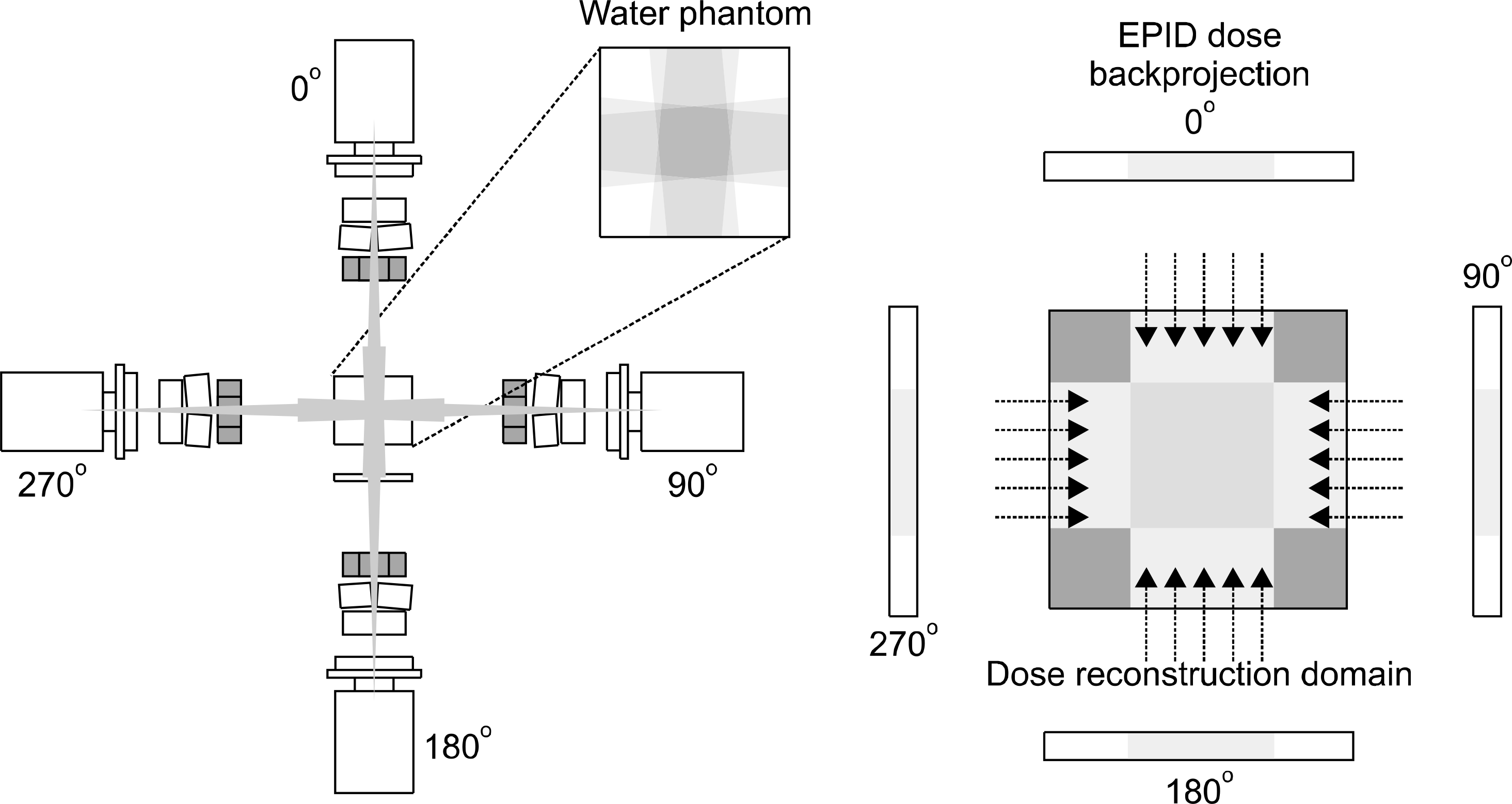
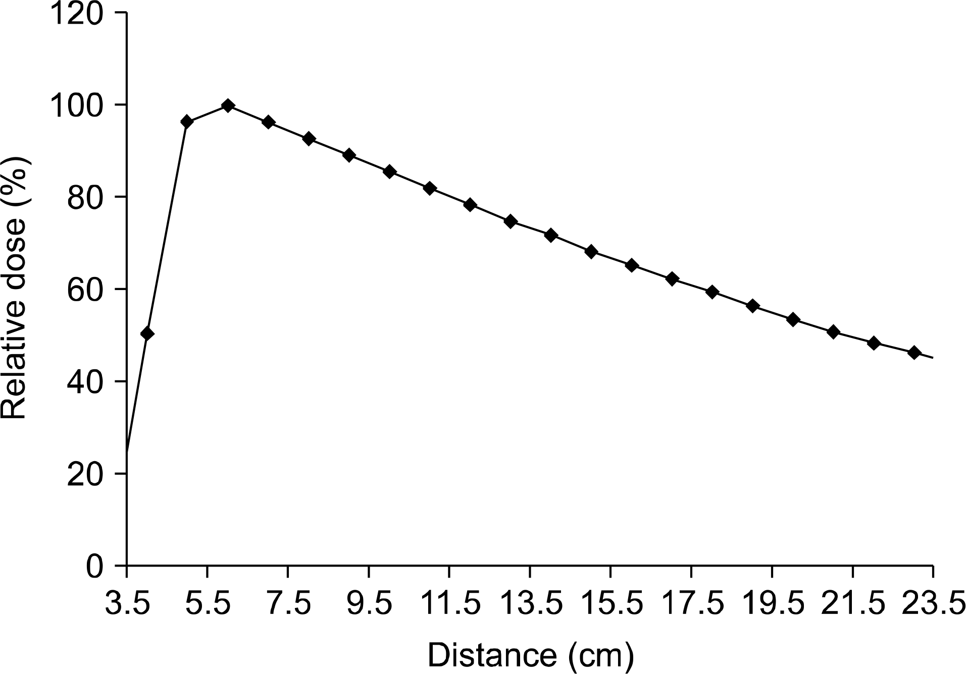
Fig. 2.
Percent depth dose (PDD) the voxel with water phantom (24.2×24.2×24.2 cm3) using 2 MeV photon source. Region of interesting set 8-16, equation of this section was adapted for calibration.
 XML Download