

 PDF
PDF ePub
ePub Citation
 Print
Print


Abstract
To investigate effects of phase mask on susceptibility-weighted images (SWI) using voxel-based analyses in normal elderly subjects. A three-dimensional (3D) gradient echo sequence ran to obtain SWIs in 20 healthy elderly subjects. SWIs with two (SWI2) and four (SWI4) phase multiplications were achieved with positive (PSWI) and negative (NSWI) phase masks to investigate phase mask effects. The voxel-based comparisons were performed using paired t-tests between PSWI and NSWI and between SWI2 and SWI4. Differences of signal intensities between magnitude images and SWI4 were larger than those between magnitude images and SWI2s. Differences of signal intensities between magnitude images and PSWIs were larger than those between magnitude images and NSWIs. Moreover, the signal intensities from NSWI2s and NSWI4s were greater than those from PSWI2s and PSWI4s, respectively. More differences of signal intensities between NSWI4 and PSWI4s were found than those between NSWI2s and PSWI2s in the whole brain images. The voxel-based analyses of SWI could be beneficial to investigate susceptibility differences on the entire brain areas. The phase masking method could be chosen to enhance brain tissue contrast rather than to enhance venous blood vessels. Therefore, it is recommended to apply voxel-based analyses of SWI to investigate clinical applications.
References
1. Haacke EM, Xu Y, Cheng YC, Reichenbach JR. Susceptibility weighted imaging (SWI). Magn Reson Med. 52(3):612–618. 2004.

2. Haacke EM, Mittal S, Wu Z, Neelavalli J, Cheng YC. Susceptibility-weighted imaging: technical aspects and clinical applications, part 1. AJNR Am J Neuroradiol. 30(1):19–30. 2009.
3. Rauscher A, Sedlacik J, Barth M, Mentzel HJ, Reichenbach JR. Magnetic susceptibility-weighted MR phase imaging of the human brain. AJNR Am J Neuroradiol. 26(4):736–742. 2005.
4. Haacke EM, Cheng NY, House MJ, et al. Imaging iron stores in the brain using magnetic resonance imaging. Magn Reson Imaging. 23(1):1–25. 2005.
5. Xu X, Wang Q, Zhang M. Age, gender, and hemispheric differences in iron deposition in the human brain: an in vivo MRI study. Neuroimage. 40(1):35–42. 2008.
6. Pfefferbaum A, Adalsteinsson E, Rohlfing T, Sullivan EV. MRI estimates of brain iron concentration in normal aging: comparison of field-dependent (FDRI) and phase (SWI) methods. Neuroimage. 47(2):493–500. 2009.
7. Haacke EM, Miao Y, Liu M, et al. Correlation of putative iron content as represented by changes in R2∗ and phase with age in deep gray matter of healthy adults. J Magn Reson Imaging. 32(3):561–576. 2010.
8. Kim MJ, Jahng GH, Lee HY, et al. Development of a Korean standard structural brain template in cognitive normals and patients with mild cognitive impairment and Alzheimer's disease. J Korean Soc Magn Reson Med. 14(2):103–114. 2010.
9. Maldjian JA, Laurienti PJ, Kraft RA, Burdette JH. An automated method for neuroanatomic and cytoarchitectonic atlas-based interrogation of fMRI data sets. Neuroimage. 19(3):1233–1239. 2003.
10. Eissa A, Lebel RM, Korzan JR, Catz I, et al. Detecting lesions in multiple sclerosis at 4.7 tesla using phase susceptibility-weighting and T2-weighting. J Magn Reson Imaging. 30(4):737–742. 2009.
11. Grabner G, Dal-Bianco A, Schernthaner M, Vass K, Lassmann H, Trattnig S. Analysis of multiple sclerosis lesions using a fusion of 3.0 T FLAIR and 7.0 T SWI phase: FLAIR SWI. J Magn Reson Imaging. 33(3):543–549. 2011.
12. Chamberlain R, Reyes D, Curran GL, et al. Comparison of amyloid plaque contrast generated by T2-weighted, T2∗-weighted, and susceptibility-weighted imaging methods in transgenic mouse models of Alzheimer's disease. Magn Reson Med. 61(5):1158–1164. 2009.
13. Niwa T, Aida N, Kawaguchi H, et al. Anatomic dependency of phase shifts in the cerebral venous system of neonates at susceptibility-weighted MRI. J Magn Reson Imaging. 34(5):1031–1036. 2011.
14. Shmueli K, de Zwart JA, van Gelderen P, Li TQ, Dodd SJ, Duyn JH. Magnetic susceptibility mapping of brain tissue in vivo using MRI phase data. Magn Reson Med. 62(6):1510–1522. 2009.
15. Schafer A, Wharton S, Gowland P, Bowtell R. Using magnetic field simulation to study susceptibility-related phase contrast in gradient echo MRI. Neuroimage. 48(1):126–137. 2009.
Fig. 1.
The representative whole brain images obtained from a healthy human subject. Magnitude (a), phase (b), positively phase-masked susceptibility-weighted image (SWI) with 2 phase mask multiplications (PSWI2, c), negatively phase-masked SWI with 2 phase mask multiplications (NSWI2, d), positively phase-masked SWI with 4 phase mask multiplications (PSWI4, e), negatively phase-masked SWI with 4 phase mask multiplications (NSWI4, f).

Fig. 2.
Results of voxel-wise comparisons between magnitude images and positively (P) phase-masked susceptibility weighted images (SWIs) with two different phase mask multiplications. Comparison between magnitude images and positively phase-masked SWI with 2 phase mask multiplications (magnitude> PSWI2, a) and between magnitude images and positively phase-masked SWI with 4 phase mask multiplications (magnitude>PSWI4, b). No significant differences were found between P_SWI2 and P_SWI4.
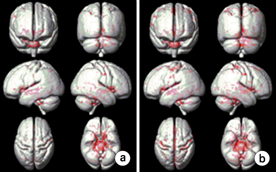
Fig. 3.
Results of voxel-wise comparisons between magnitude images and negatively (N) phase- masked susceptibility weighted image (SWI) with two different phase mask multiplications. Comparisons between magnitude images and negatively phase-masked SWI with 2 phase mask multiplications (magnitude>NSWI2, (a), between magnitude images and negatively phase-masked SWI with 4 phase mask multiplications (magnitude>NSWI4, (b), and between NSWI2 and NSWI4 (NSWI2>NSWI4, c).
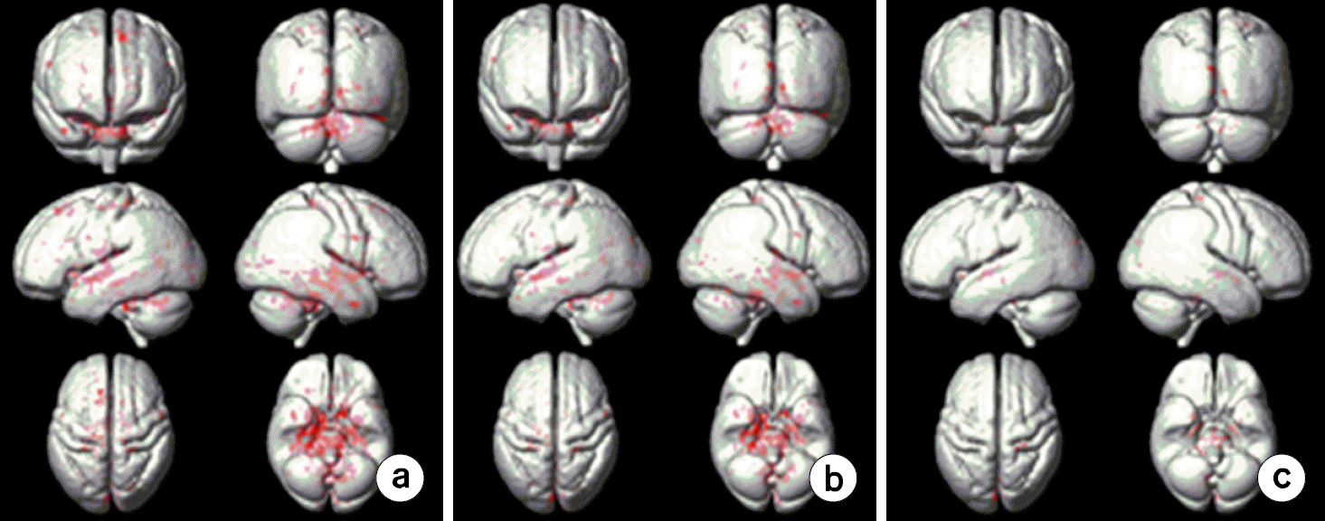
Fig. 4.
Results of voxel-wise comparisons between susceptibility-weighted imaging (SWI) with the positive (P) phase mask and that with the negative (N) phase mask with the whole brain with the threshold of family-wise error rate (FWE) p=0.005. (a) Comparison between the whole brain negatively phase-masked SWI with 2 phase mask multiplications and positively phase-masked SWI with 2 phase mask multiplications (NSWI2> PSWI2). (b) Comparison between the whole brain negatively phase-masked SWI with 4 phase mask multiplications and positively phase-masked SWI with 4 phase mask multiplications (NSWI4>PSWI4).
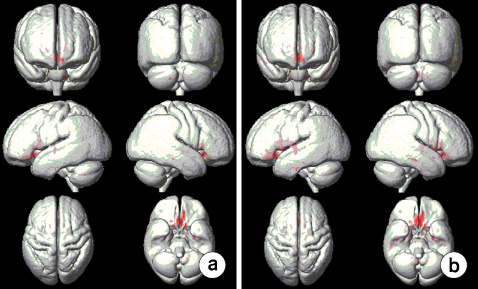
Table 1.
Anatomical regions showing significant differences between magnitude and positively (P) phase-masked susceptibility-weighted imaging (SWI).
Table 2.
Anatomical regions showing significant differences between magnitude and negatively (N) phase-masked susceptibility-weighted imaging (SWI).
Table 3.
Anatomical regions showing significant differences between the whole brain susceptibility-weighted imaging (SWI) with the positive (P) phase mask and that with the negative (N) phase mask.
 XML Download