

 PDF
PDF ePub
ePub Citation
 Print
Print


Abstract
The aim of this study was to clarify the impacts of acquisition parameters on artifacts in four-dimensional computed tomography (4D CT) images, such as the partial volume effect (PVE), partial projection effect (PPE), and mis-matching of initial motion phases between adjacent beds (MMimph) in cine mode scanning. A thoracic phantom and two cylindrical phantoms (2 cm diameter and heights of 0.5 cm for No.1 and 10 cm for No.2) were scanned using 4D CT. For the thoracic phantom, acquisition was started automatically in the first scan with 5 sec and 8 sec of gantry rotation, thereby allowing a different phase at the initial projection of each bed. In the second scan, the initial projection at each bed was manually synchronized with the inhalation phase to minimize the MMimph. The third scan was intentionally un-synchronized with the inhalation phase. In the cylindrical phantom scan, one bed (2 cm) and three beds (6 cm) were used for 2 and 6 sec motion periods. Measured target volume to true volume ratios (MsTrueV) were computed. The relationships among MMimph, MsTrueV, and velocity were investigated. In the thoracic phantom, shorter gantry rotation provided more precise volume and was highly correlated with velocity when MMimph was minimal. MMimph reduced the correlation. For moving cylinder No. 1, MsTrueV was correlated with velocity, but the larger MMimph for 2 sec of motion removed the correlation. The volume of No. 2 was similar to the static volume due to the small PVE, PPE, and MMimph. Smaller target velocity and faster gantry rotation resulted in a more accurate volume description. The MMimph was the main parameter weakening the correlation between MsTrueV and velocity. Without reducing the MMimph, controlling target velocity and gantry rotation will not guarantee accurate image presentation given current 4D CT technology.
REFERENCES
1. Lecchi M, Fossati P, Elisei F, Orecchia R, Lucignani G. Current concepts on imaging in radiotherapy. Eur J Nucl Med Mol Imaging. 35(4):821–837. 2008.

2. Rietzel E, Pan T, Chen GT. Four-dimensional computed tomography: image formation and clinical protocol. Med Phys. 32(4):874–889. 2005.
3. Wong JW, Sharpe MB, Jaffray DA, et al. The use of active breathing control (ABC) to reduce margin for breathing motion. Int J Radiat Oncol Biol Physics. 44:911–919. 1999.
4. Hanley J, Debois NM, Mah D, et al. Deep inspiration Breath-hold technique for lung tumors: the potential value of target immobilization and reduced lung density in dose escalation: Int J Radiat Oncol Biol Physics. 45:603–611. 1999.
5. Shin EH, Park HC, Han YY, et al. Efficacy of a respiratory training system on the regularity of breathing. J. Korean Soc Ther Radiol. 26(3):181–188. 2008.
6. Minohara S, Endo M, Kanai T, Kato H, Tsujii H. Estimating uncertainties of the geometrical range of particle radiotherapy during respiration. Int J Radiat Oncol Biol Phys. 56(1):121–125. 2003.
7. Wurstbauer K, Deutschmann H, Kopp P, Sedlmayer F. Radiotherapy planning for lung cancer: slow CTs allow the drawing of tighter margins. Radiother Oncol. 75(2):165–170. 2005.
8. Lagerwaard FJ, Van Sornsen de Koste Jr, Nijssen- Visser MR, et al. Multiple "slow" CT scans for incorporating lung tumor mobility in radiotherapy planning. Int J Radiat Oncol Biol Phys. 51(4):932–937. 2001.
9. Vedam SS, Keall PJ, Kini VR, Mostafavi H, Shukla HP, Mohan R. Acquiring a four-dimensional computed tomography dataset using an external respiratory signal. Phys Med Biol. 48(1):45–62. 2003.
10. Keall PJ. 4-dimensional computed tomography imaging and treatment planning. Semin Radiat Oncol. 14(1):81–90. 2004.
11. Keall PJ, Starkschall G, Shukla H, et al. Acquiring 4D thoracic CT scans using a multislice helical method. Phys Med Biol. 49(10):2053–2067. 2004.
12. Mori S, Endo M, Asakura H. Improvement in banding artifacts in four-dimensional computed tomography for radiotherapy planning. Phys Med Biol. 51(20):5231–5244. 2006.
13. Nakamura M, Narita Y, Sawada A, et al. Impact of motion velocity on four-dimensional target volumes: a phantom study. Med Phys. 36(5):1610–1617. 2009.
14. Pan T. Comparison of helical and cine acquisitions for 4D-CT imaging with multislice CT. Med Phys. 32(2):627–634. 2005.
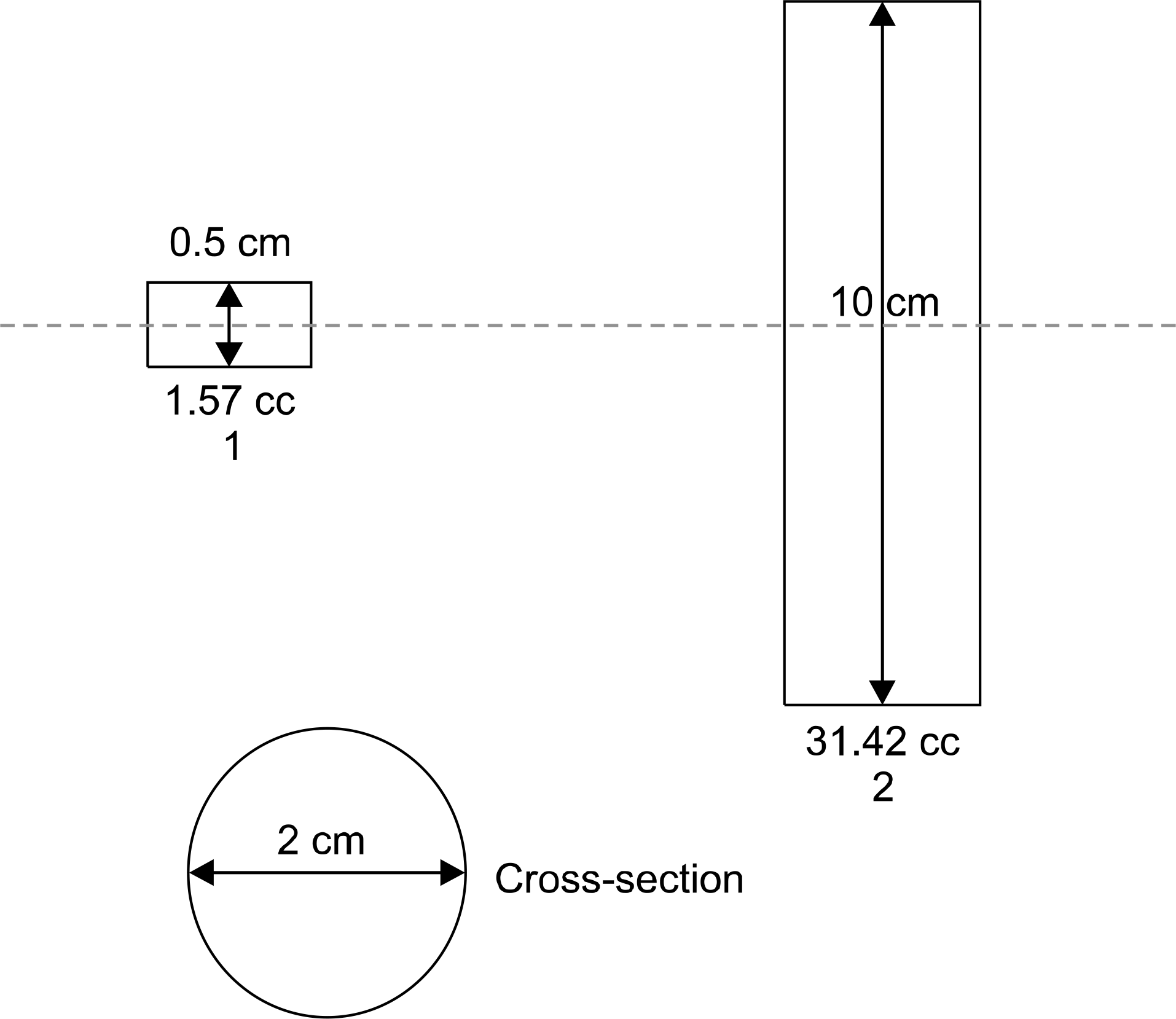
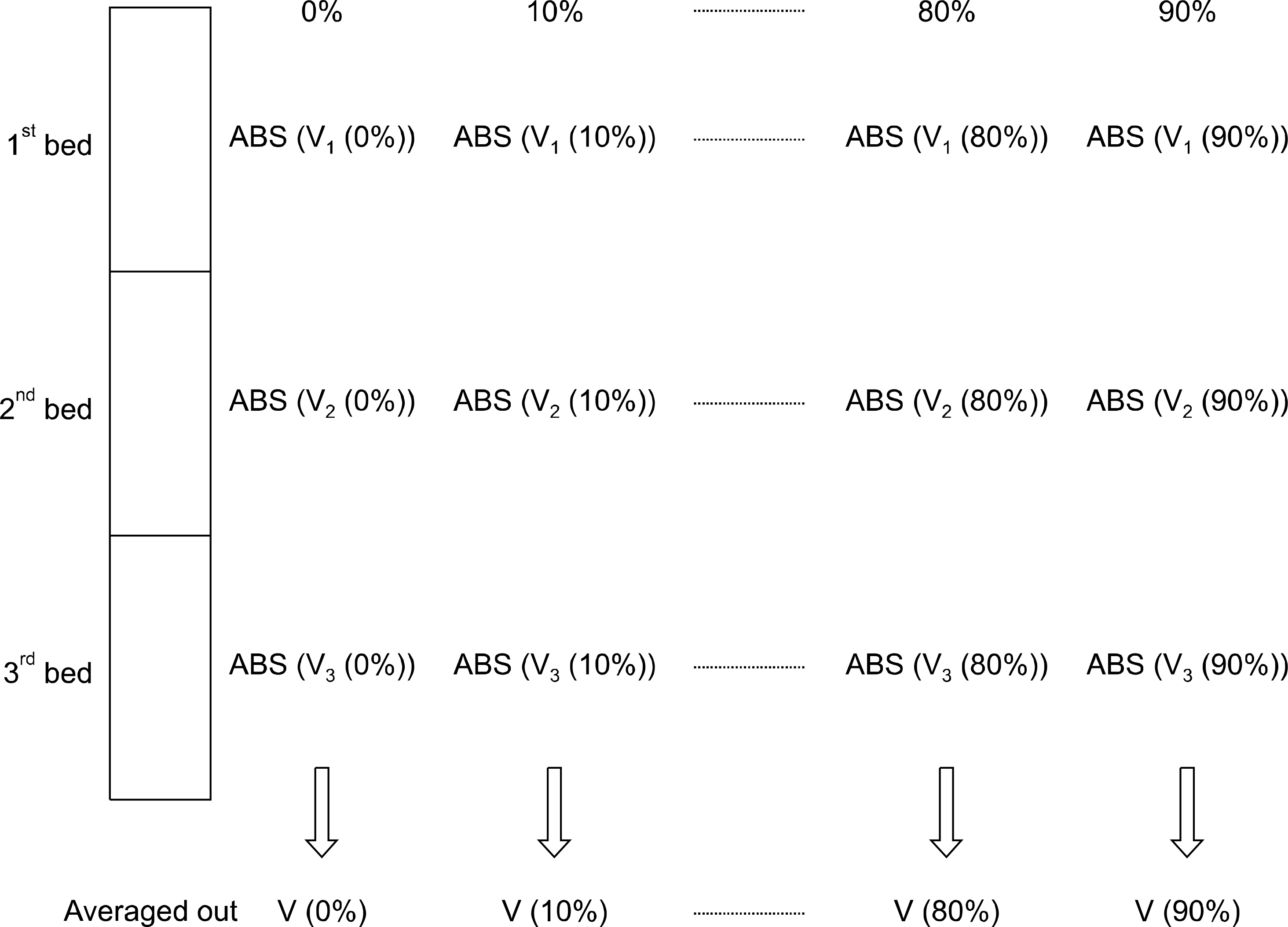
Fig. 3.
The correlation between ABS(Vdifference (%)) and the correlation between MsTrueV and velocity in the moving thoracic phantom.
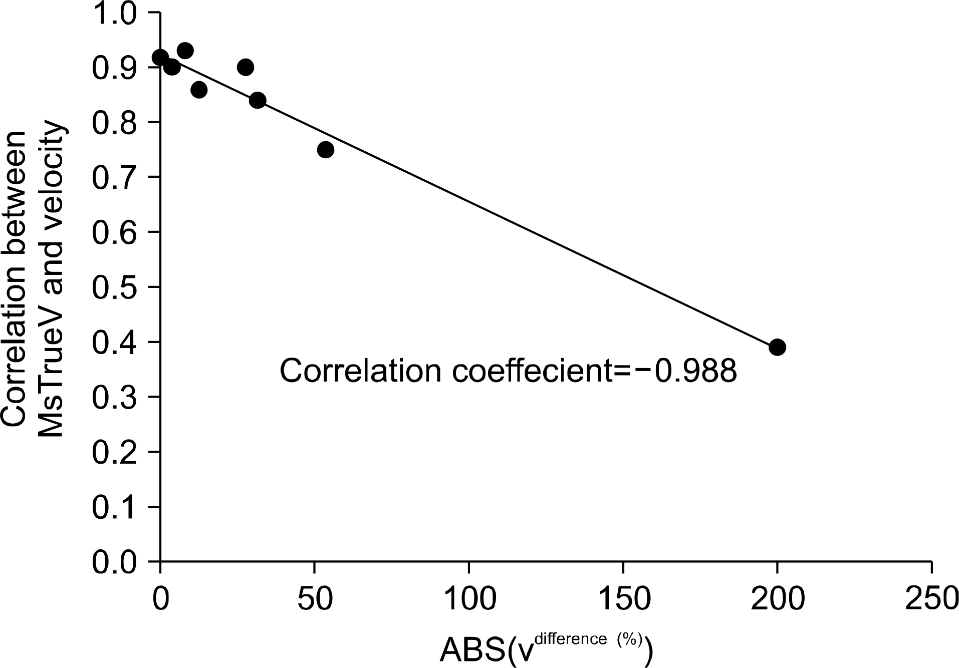
Fig. 4.
Velocity and MsTrueV values obtained from 1A4P (amplitude 1 cm, period 4 sec), 1A6P, 2A4P, and 2A6P using the thoracic phantom. The scan was automatically started, and the image slice thickness was 2.5 mm.
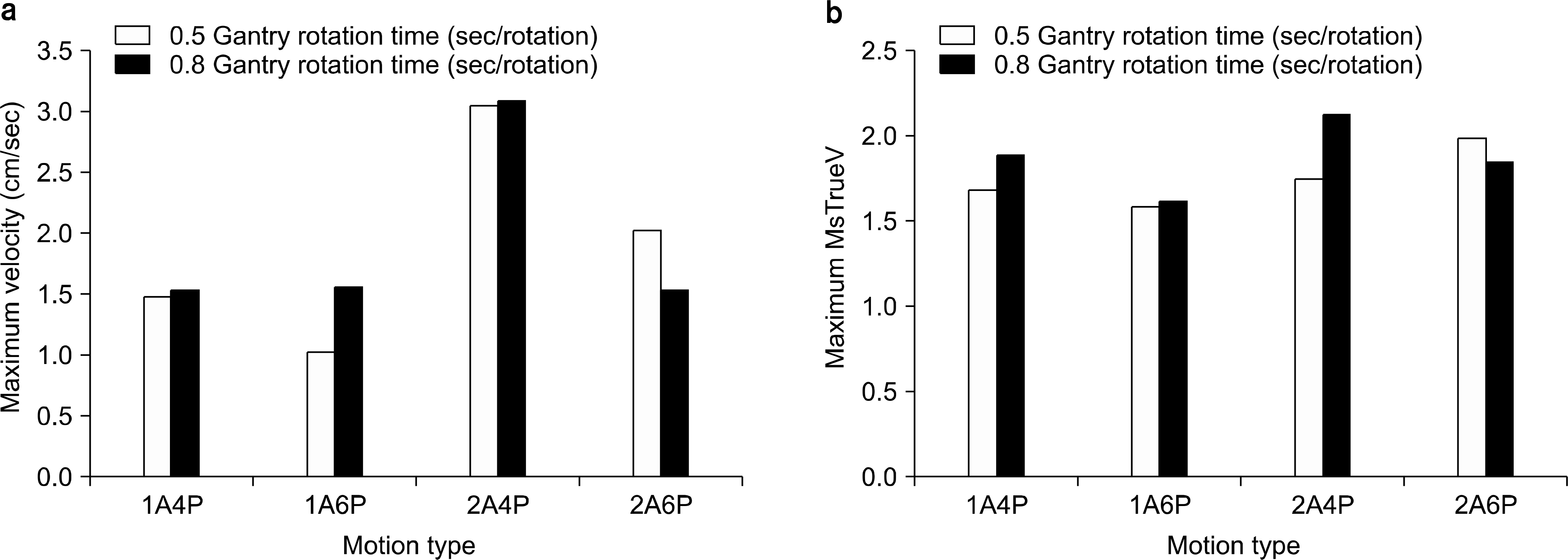
Fig. 5.
4D CT motion artifacts in 1A6P (amplitude 1 cm, period 6 sec) and 2A4P with 0.8 sec gantry rotation time: a) 0% of respiratory phase for 1A6P, b) 30% of respiratory phase for 1A6P, c) 0% of respiratory phase for 2A4P, and d) 30% of respiratory phase for 2A4P.
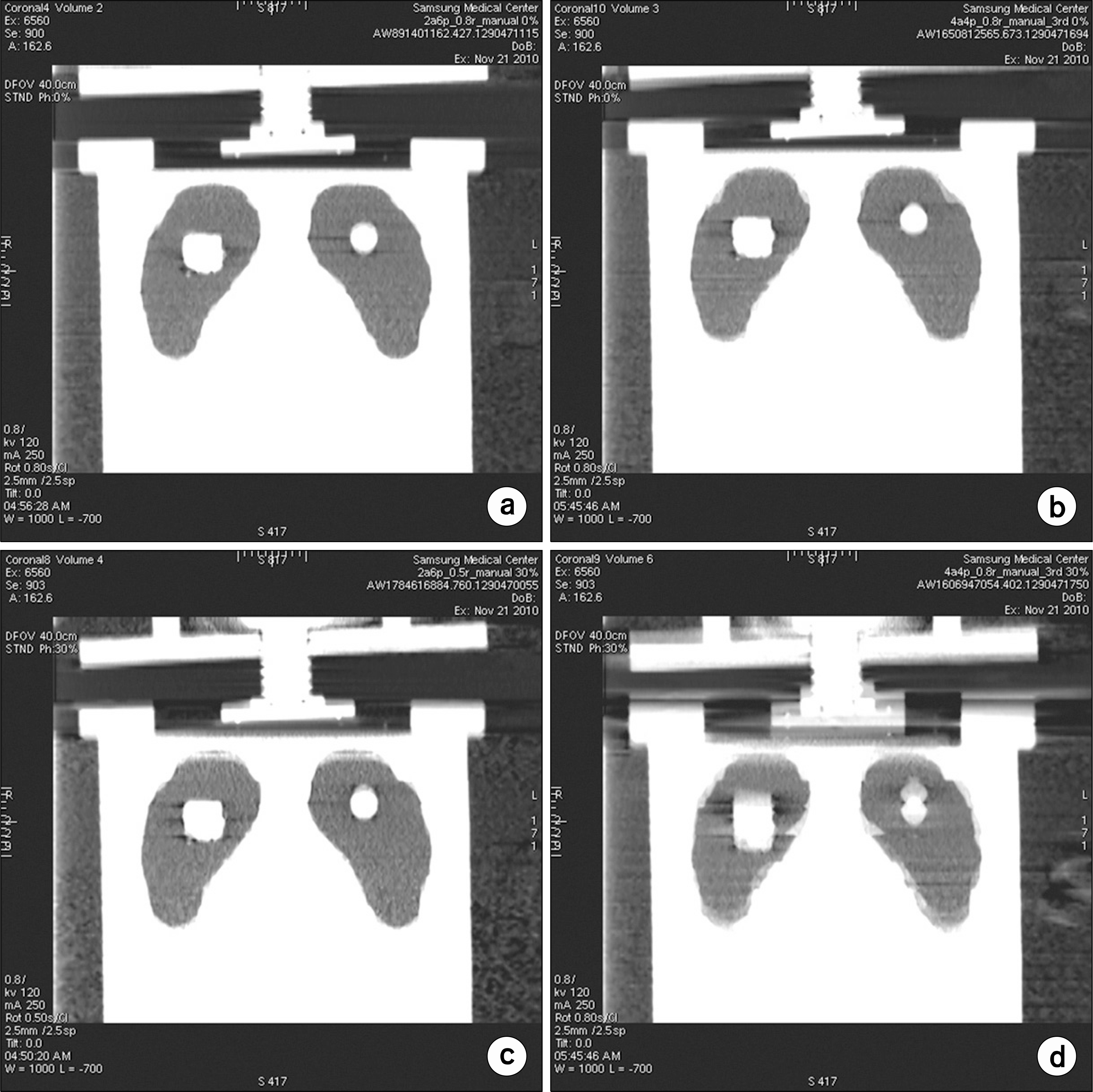
Fig. 6.
Calculated velocity vs. percentage phase actually detected by RPM at each bed. The first phase at the X-axis indicates the phase at gantry angle 0. Phantom motion had an amplitude of 0.5 cm and a motion period 4 sec. 1A4P: gantry rotation time 0.8 sec, interleaved between images for 0.4 sec in three beds. In the first bed, the initial phase at a gantry angle of 0 degrees was 40% (actual 41%), while the initial phases changed to 0% and 50% in the second and third beds, respectively. The different initial projection angles of the phantom, which caused large ABS(Vdifference (%)), resulted in different velocities at the same nominal phase percent range among beds, which were 41%, 35%, and 37% in the first, second, and third beds, respectively, for the nominal 40% phase.
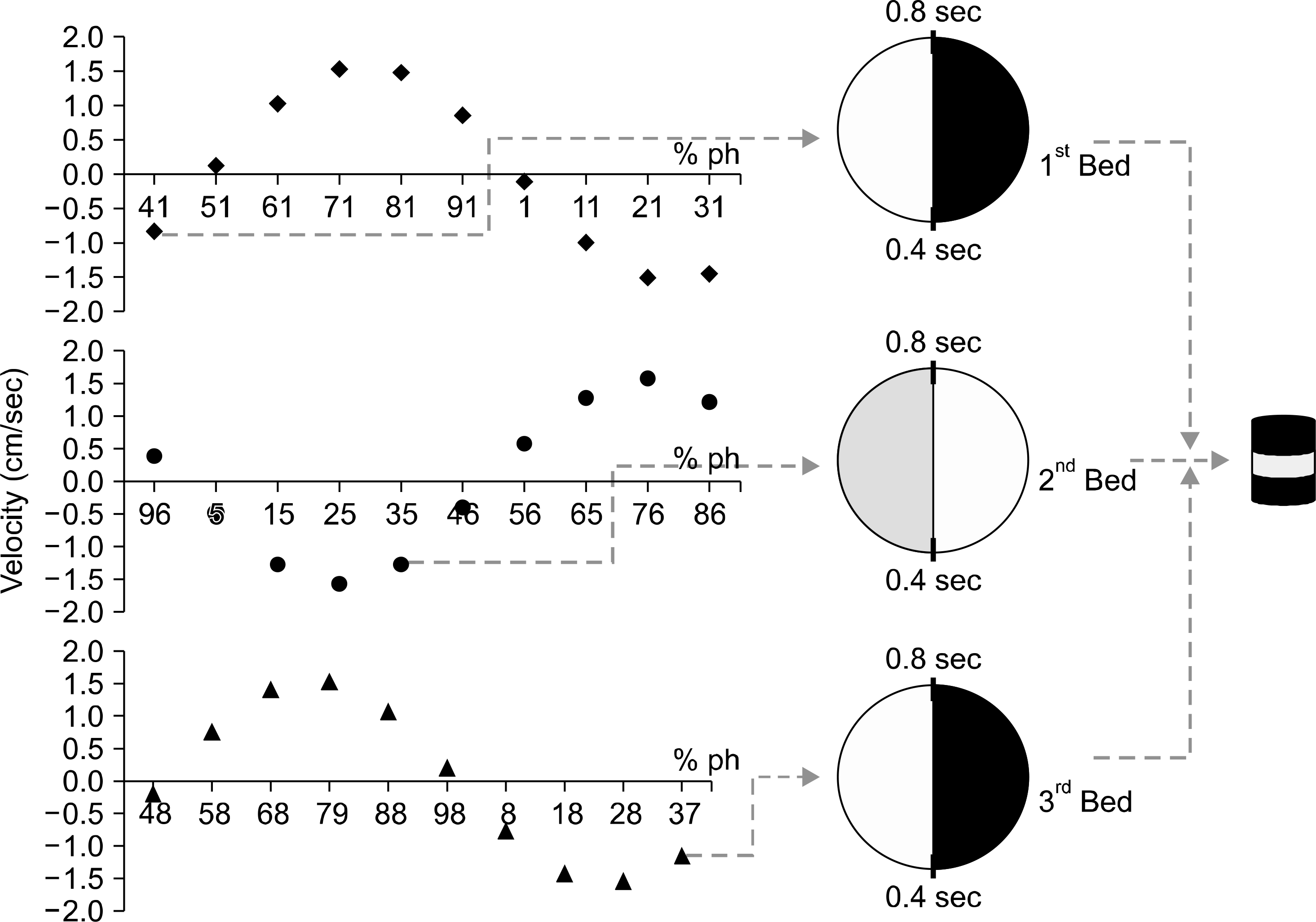
Table 1.
Velocity and MsTrueV values obtained from 1A4P (amplitude 1 cm, period 4 sec), 1A6P, 2A4P, and 2A6P using the thoracic phantom. The scan was automatically started, and the image slice thickness was 2.5 mm.
Table 2.
MMimph and MsTrueV: MsTrueV for 2 cm amplitude with 4 sec and 6 sec period motion (2A4P and 2A6P), 1A4P, and 1A6P. Gantry rotations were 0.8 sec per rotation. Images slice thickness was 2.5 mm. MMimph: Vj(p) is velocity of p% phase in the jth bed (A: amplitude, p: % phase). ABS(Vj(p))=|(Vj(p)|, Vdifference (%)={ABS(Vj-1(p')–ABS (Vj(p)}∗100(%)/ABS (Vj-1(p)).
Table 3.
Acquisition parameters, volume, and MsTrueV values for the static cylindrical phantom.
Table 4.
MsTrueV values for cylindrical phantoms moving with 0.5 cm amplitude and a 6 sec cycle.
Table 5.
Computed velocity differences at the starting projection for each bed and MsTrueV values for the cylindrical phantoms with 0.5 cm amplitude and 6 sec or 2 sec of motion for three-bed scanning.
 XML Download