

 PDF
PDF ePub
ePub Citation
 Print
Print


Abstract
Upon radiation treatment, it is the important factor to monitor the patient's motion during radiation irradiated, since it can determine whether the treatment is successful. Thus, we have developed the system in which the patient's motion is monitored in real time and moving treatment position can be automatically corrected during radiation irradiation. We have developed the patient's position monitoring system in which the patient's position is three dimensionally identified by using two CCD cameras which are orthogonal located around the isocenter. This system uses the image pattern matching technique using a normalized cross-correlation method. We have developed the system in which trigger signal for beam on and off is generated by quantitatively analyzing the changes in a treatment position through delivery of the images taken from CCD cameras to the computer and the motor of moving couch can be controlled. This system was able to automatically correct a patient's position with the resolution of 0.5 mm or less.
Go to : 
REFERENCES
1. Cho BC, Huh HD, Kim JS, et al. Guideline for imaging dose on image-guided radiation therapy. Progress in Medical Physics. 24(1):1–24. 2013.

2. ICRU Report 62. Prescribing Recording and Reporting Photon Beam Therapy (supplement to ICRU Report 50). International Commission on Radiation Units and Measurements, Bethesda, MD. 1999.
3. Kwon KT, Lim SW, Park SH, et al. Evaluation of difference between skin motion and tumor motion for respiration gated radiotherapy. Progress in Medical Physics. 19(1):14–20. 2008.
4. Huh HD, Choi SH, Kim WC, et al. Analysis of dose distribution on critical organs for radiosurgery with cyberknife real- time tumor tracking system. Progress in Medical Physics. 20(1):14–20. 2009.
5. Seo JH, Kang YN, Jang JS, et al. Estimation of cyberknife respiratory tracking system using moving phantom. Progress in Medical Physics. 20(4):324–330. 2009.
6. Hsi WC, Moyers MF, Nichiporov D, et al. Energy spectrum control for modulated proton beams. Medical Physics. 36(6):2297–2308. 2009.
7. Anferov VA. Scan pattern optimization for uniform proton beam scanning. Medical Physics. 36(8):3560–3567. 2009.
8. Farr JB, Dessy F, De Wilde O, et al. Fundamental radiological and geometric performance of two types of proton beam modulated discrete scanning systems. Medical Physics. 40(7):07210–1. -07210-8. 2013.
9. NI Vision Concepts Manual. National Instrument, Austin Texas (2005). pp. 12-7-8.
10. Rogus RD, Stern RL, Kubo HD. Accuracy of a photogrammetry- based patient positioning and monitoring system for radiation therapy. Medical Physics. 26(5):721–728. 1999.
11. Kim HY, Park YK, Kim IH, et al. Development of an optical- based image guidance system: Technique detecting ex ternal markers behind a full facemask. Medical Physics. 38(6):3006–3012. 2011.
12. Schewe JE, Lam KL, Balter JM, et al. A room-based diagnostic imaging system for measurement of patient setup. Medical Physics. 25(12):2385–2387. 1998.
Go to :
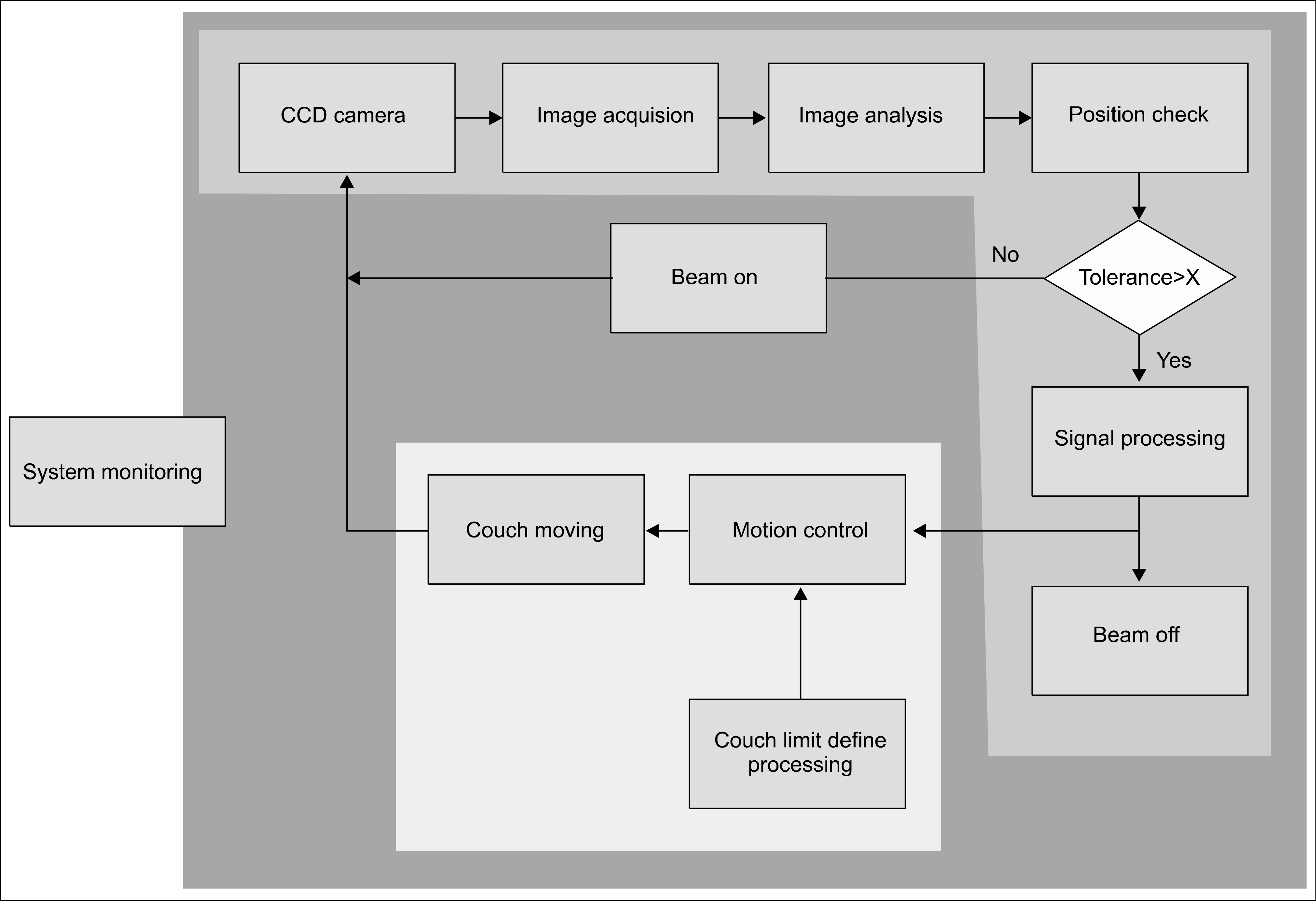 | Fig. 1.Schematic diagram for algorithm of automatic patient position correction system software and hardware. |
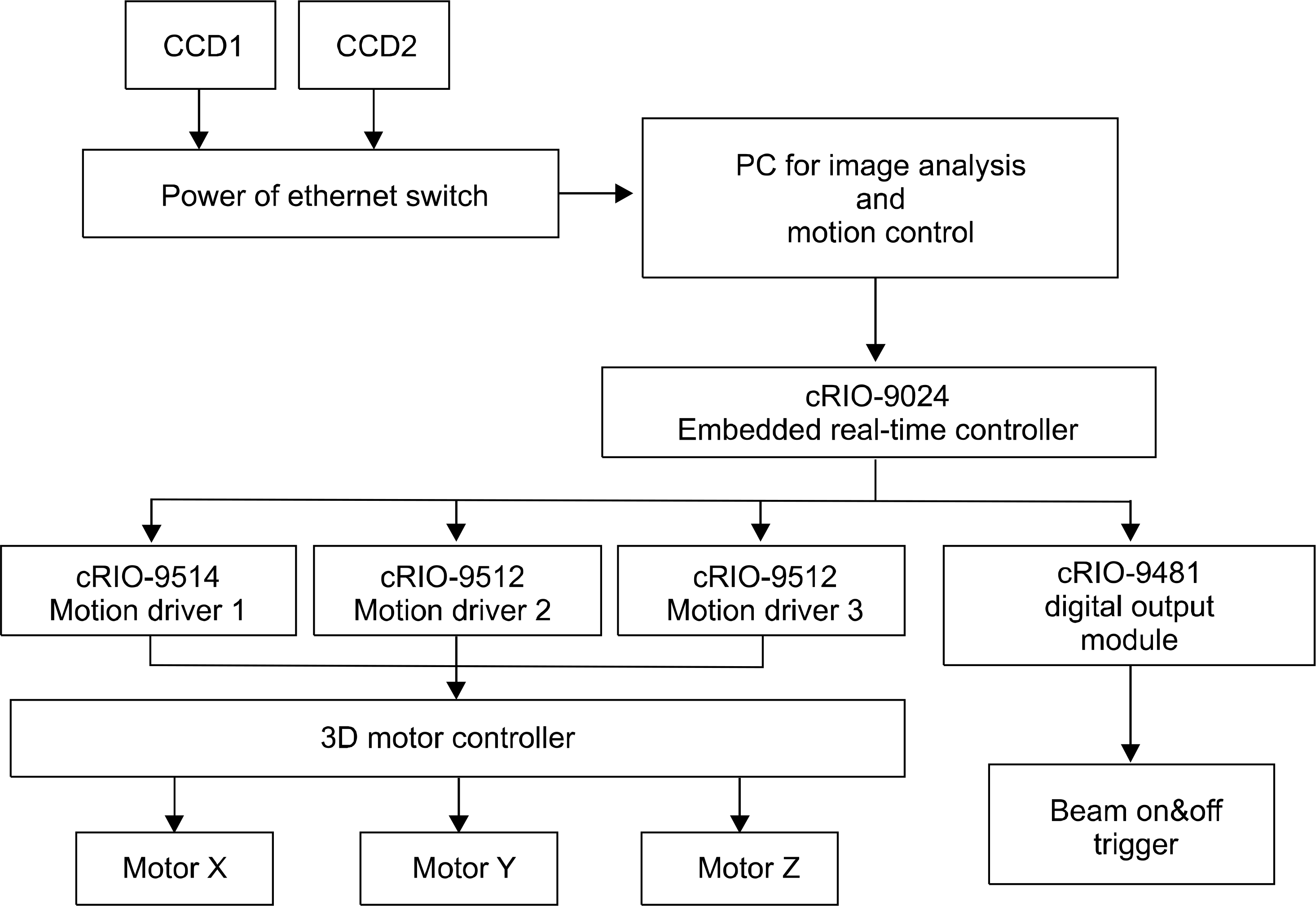 | Fig. 2.Schematic diagram for H/W connection for automatic patient position correction system. |
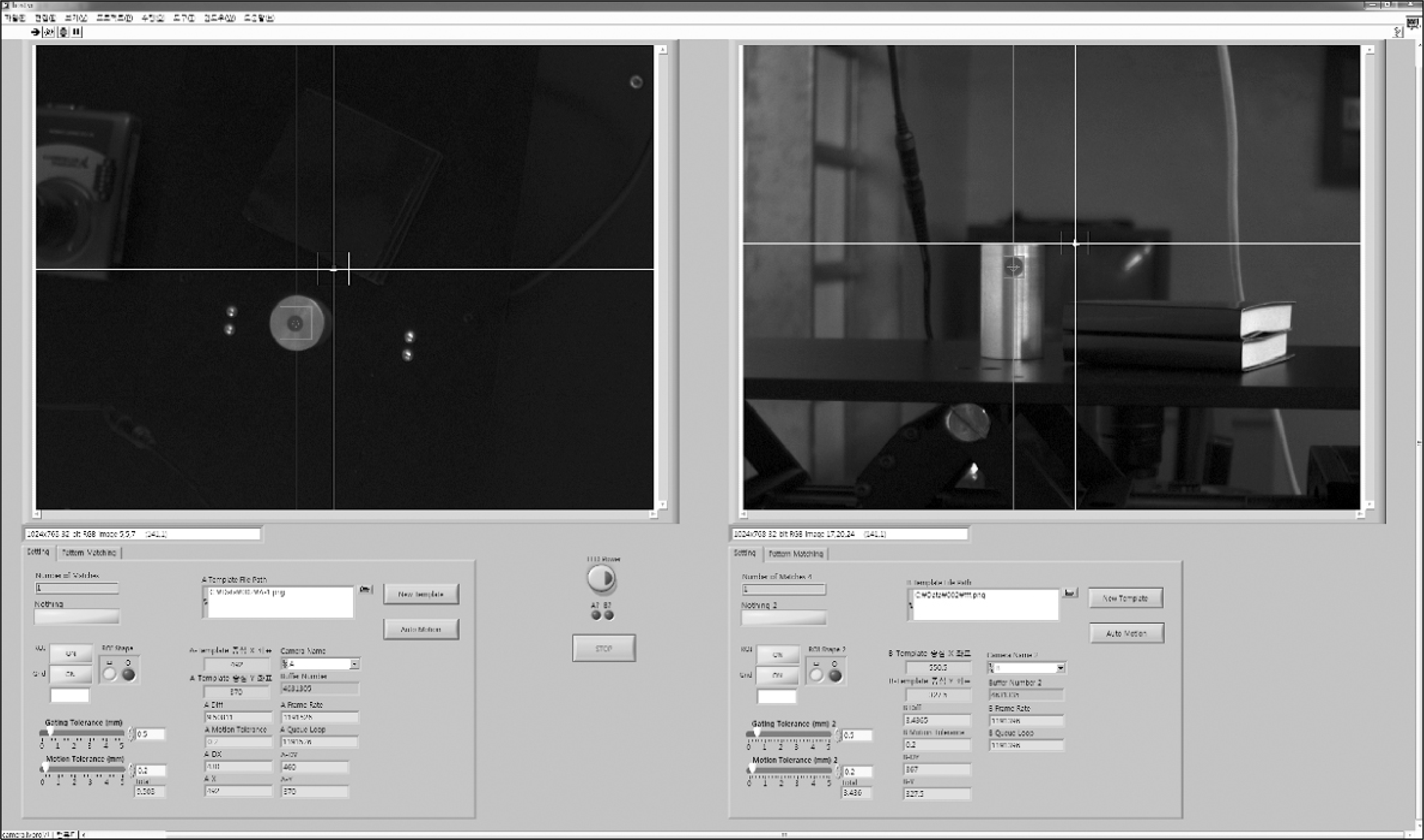
 | Fig. 3.Pattern matching example. (left) pattern matching circle point marker for x-y plane, (right) pattern matching circle point marker for z-axis. |
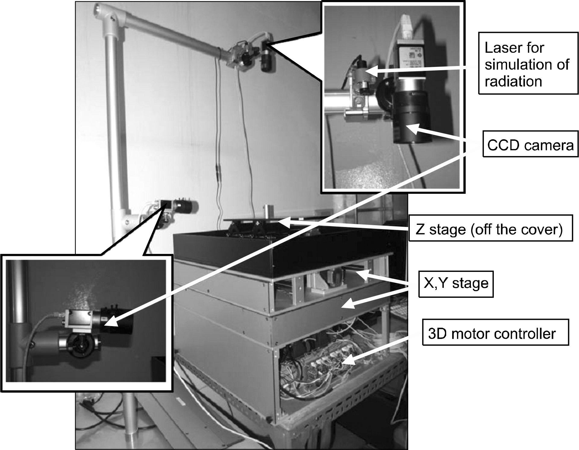
 | Fig. 4.The home made 3D moving phantom for automatic position correction system and CCD camera holder. |
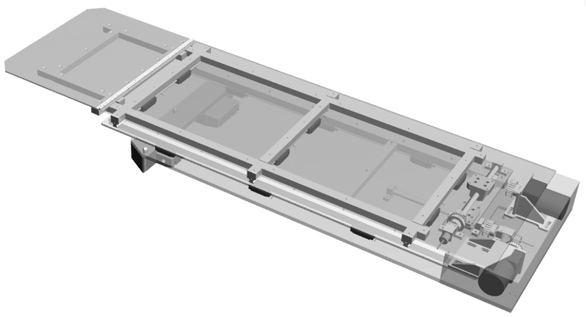 | Fig. 5.Schematic diagram for 3D modeling of the 3D moving couch for heavy moving phantom motion correction verification. |
Table 1.
The result of rectal cancer patient treatment setup position difference of between before treatment and after treatment.
 XML Download