

 PDF
PDF ePub
ePub Citation
 Print
Print


Abstract
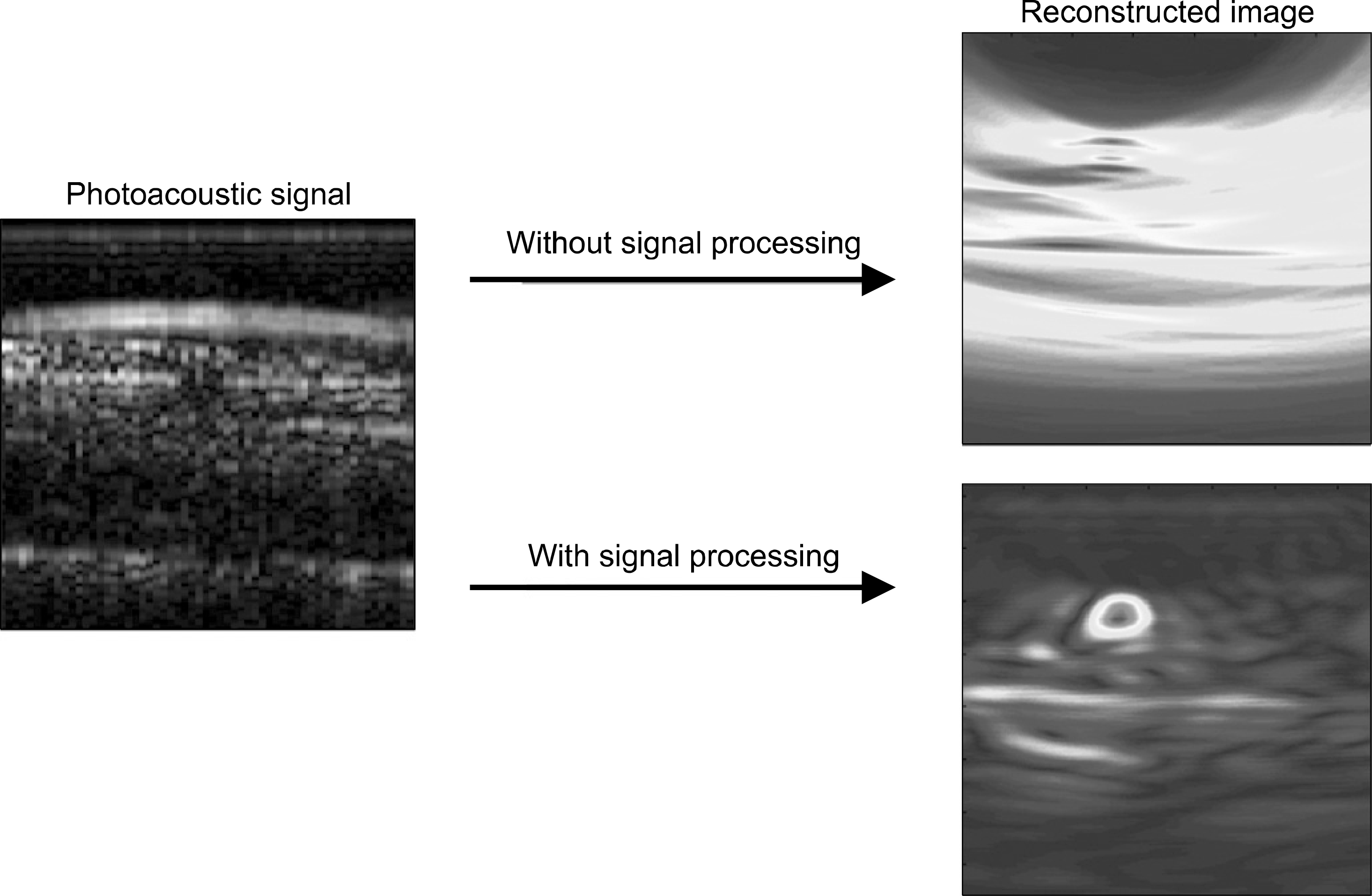
Recently, the photoacoustic imaging system has been widely and intensively developed, and has been shown the possibility of diagnosis for early stage cancer. In this study, we developed a photoacoustic tomography imaging system with a commercial ultra sound device and a linear array probe. A tube phantom and a chicken breast phantom was made for the possibility of a system as a breast cancer detection. A moving average filter and a band pass filter with 3∼6 MHz bandwidth were developed for background noise elimination before delay-and-sum beamforming algorithm was used for image reconstruction. As a result, we showed that some signal processing procedure before beamforming was effective for the photoacoustic image reconstruction.
REFERENCES
1. Xu M, Wang LH. Photoacoustic imaging in biomedicine. Review of Scientific Instruments. 77(4):041101. 2006.

2. Li C, Wang LH. Photoacoustic tomography and sensing in biomedicine Phys Med Biol. 54(19):59–97. 2009.
3. Weidner N, Semple JP, Welch WR, Folkman J. Tumor angiogenesis and metastasis–correlation in invasive breast carcinoma. N Engl J Med. 324(1):1–8. 1991.
4. Heijblom M, Piras D, Xia W, et al. Visualizing breast cancer using the Twente photoacoustic mammoscope: What do we learn from twelve new patient measurements? Opt Express. 20(11):11582–11597. 2012.
5. 김주혜, 허장용, 오정환 등. 유방암 진단용 광음향 영상 시스템 특성 평가를 위항 팬텀 개발. 의학물리. 23(1):28–30. 2012.
6. Park SH, Aglyamov SR, Emelianov SY. Beamforming for photoacoustic imaging using linear array transducer. IEEE Ultrasonics Symposium 856-859. 2007.
7. Kruger RA, Kiser WL, Reinecke DR, Kruger GA. Thermoacoustic computed tomography using a conventional linear transducer array. Medical Physics. 30(5):856–860. 2003.
8. Ku G, Wang X, Stoica G, Wang LH. Multiple-bandwidth photoacoustic tomography. Phys Med Biol. 49(7):1329–38. 2004.
9. Duck FA. Physical Properties of Tissue. Academic Press;London: 1990. ), pp.p. 120–130.
10. Wells PNT. Ultrasonic imaging of the human body. Rep Prog Phys. 62(5):671. 1999.
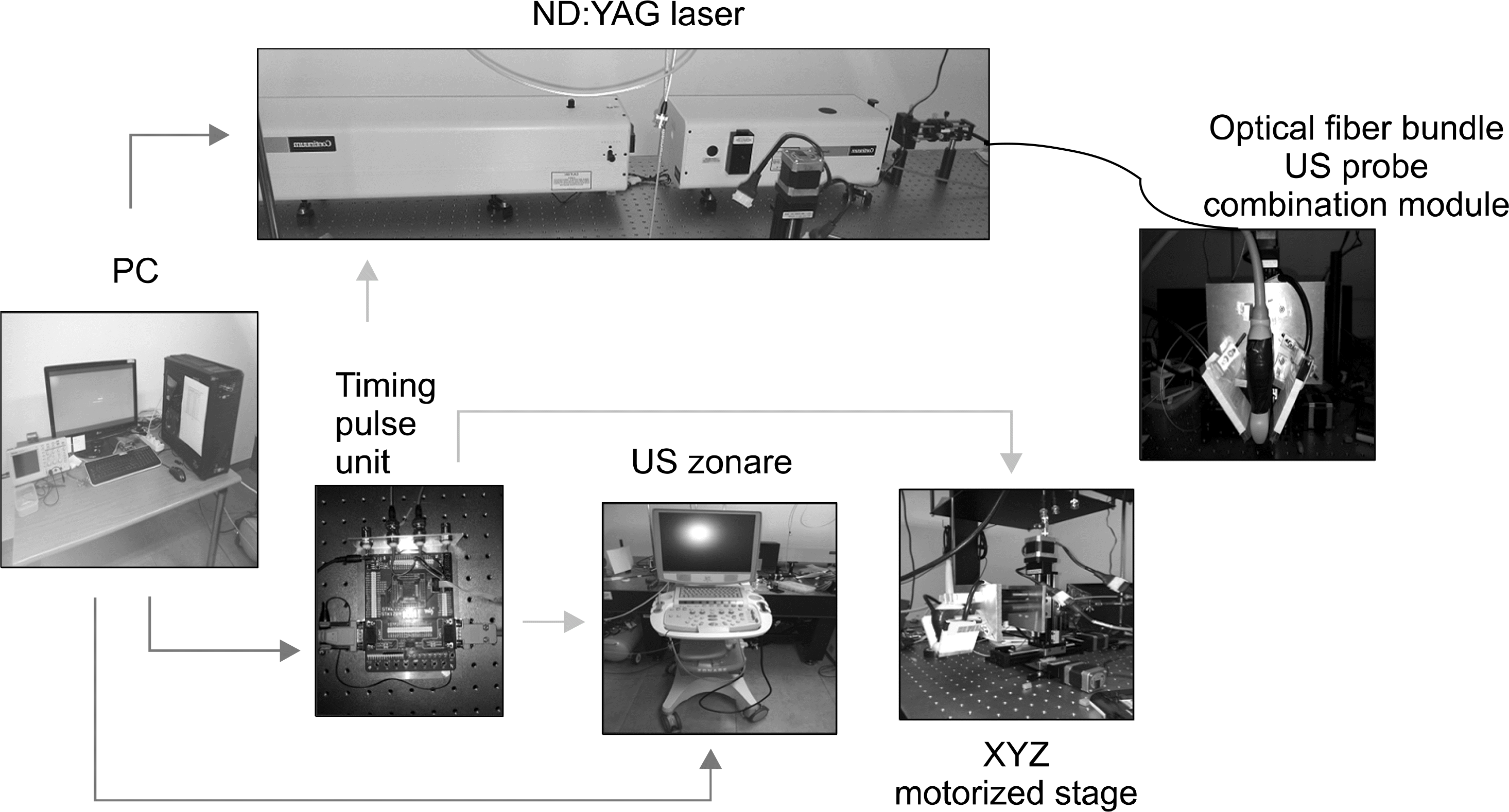
Fig. 2.
(a) Silicon tube phantom filled with methylene blue, (b) Chicken breast phantom inserted with silicon tube filled with methylene blue, (c) Measurement of tube phantom by probe module, (d) Measurement of chicken breast phantom by probe module.
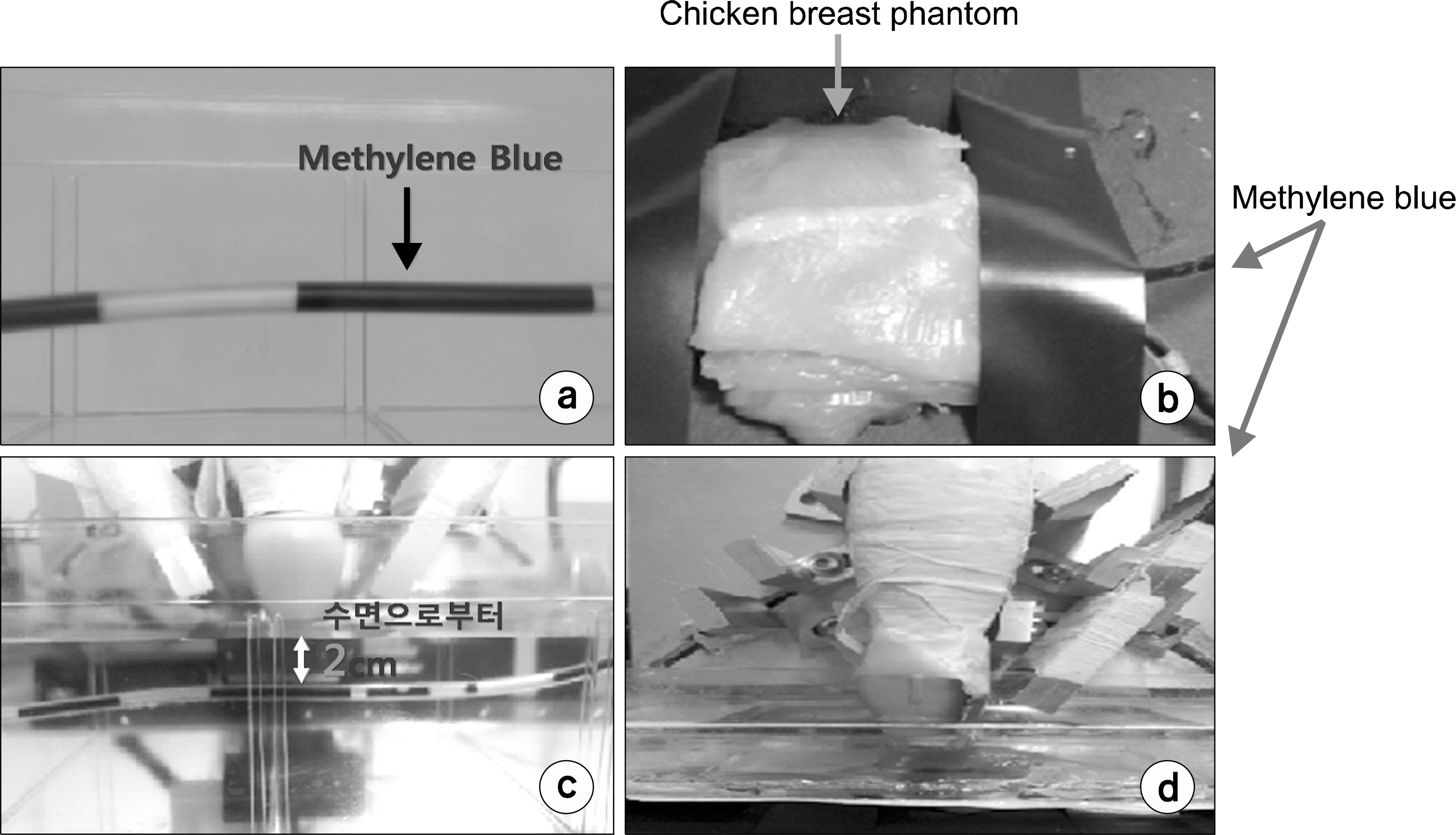
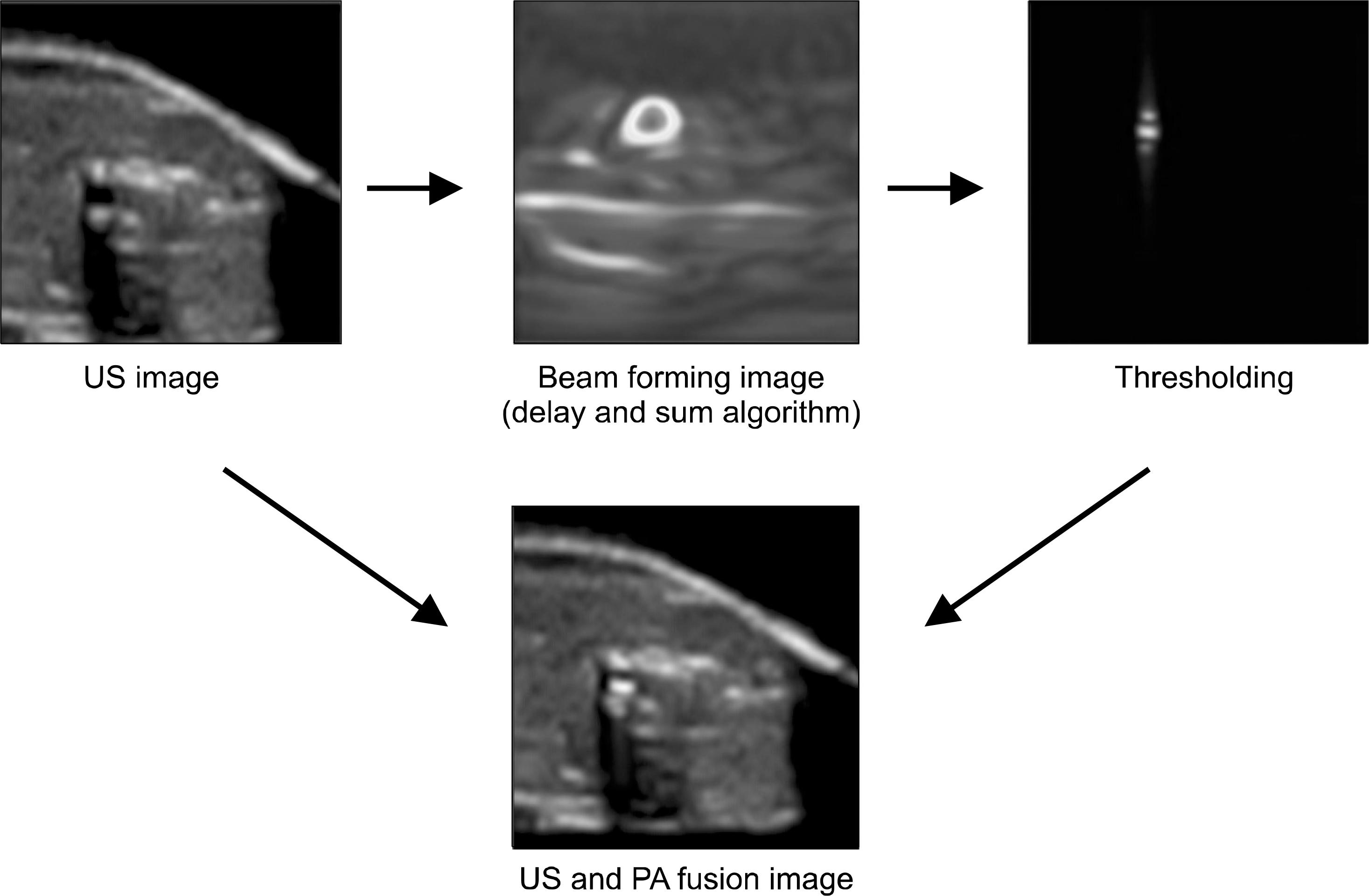
Fig. 3.
(a) Ultra sound image of tube phantom, (b) Photoacoustic signal of tube phantom, (c) Reconstructed image by DAS algorithm, (d) XY plane reconstructed image by 3D scanning.
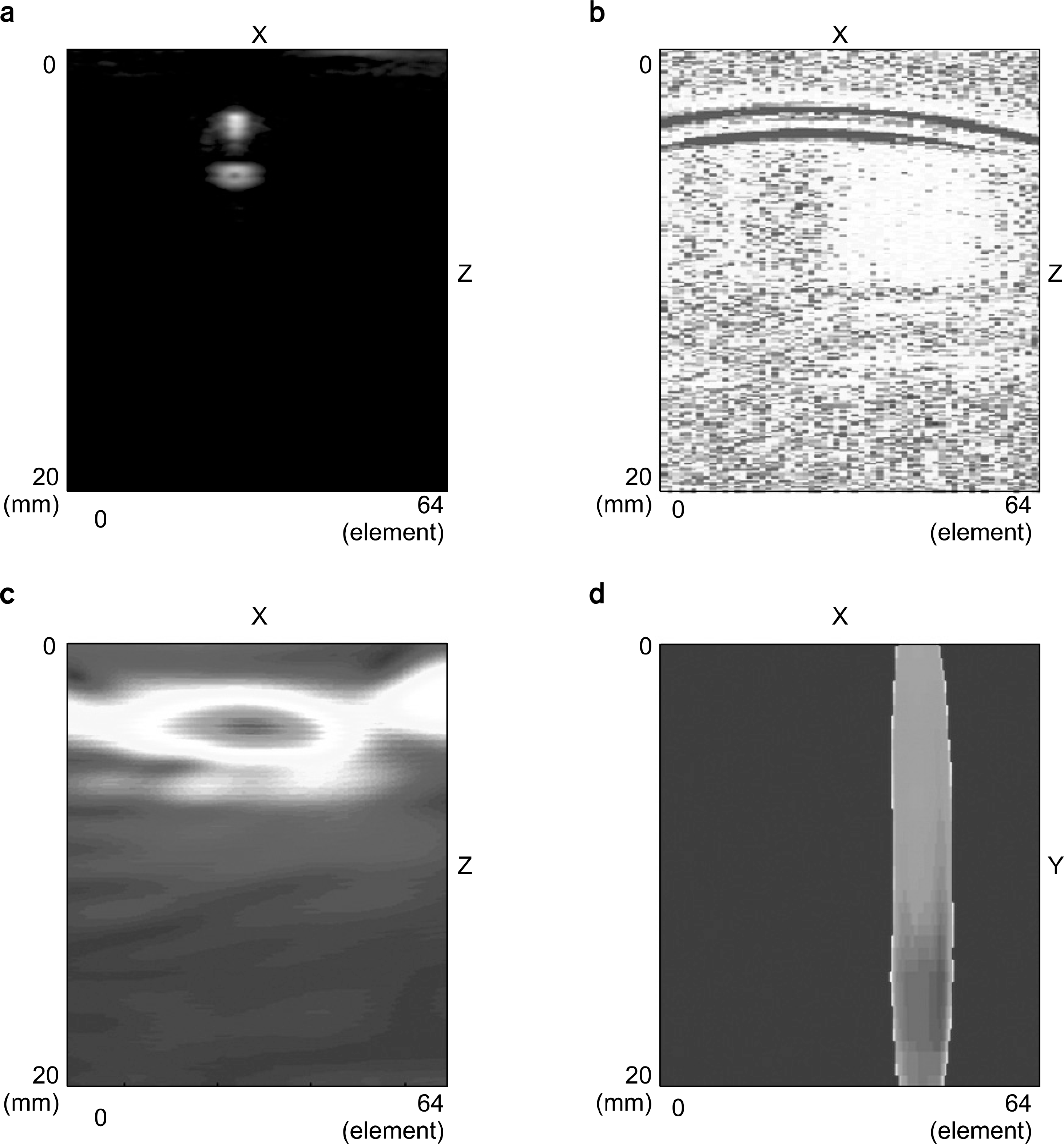
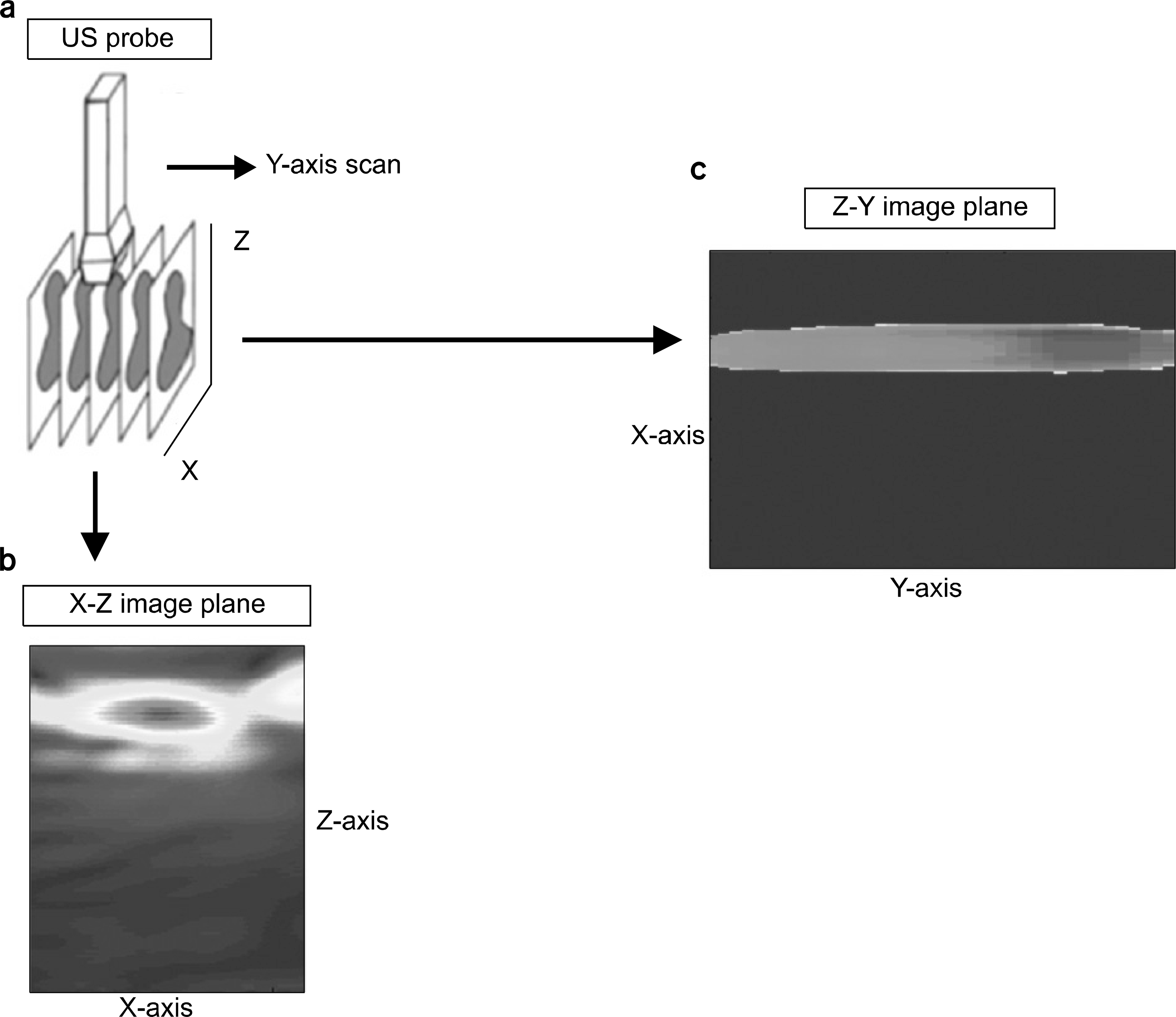
Fig. 4.
3D scanning method. (a) Y-axis scanning. (b) Photoacoustic image of x-z image. (c) Reconstructed x-y plane image of a fixed z-depth.
 XML Download