Abstract
The position of the internal organs can change continually and periodically inside the body due to the respiration. To reduce the respiration induced uncertainty of dose localization, one can use a respiratory gated radiotherapy where a radiation beam is exposed during the specific time of period. The main disadvantage of this method is that it usually requests a long treatment time, the massive effort during the treatment and the limitation of the patient selection. In this sense, the combination of the real-time position management (RPM) system and the volumetric intensity modulated radiotherapy (RapidArc) is promising since it provides a short treatment time compared with the conventional respiratory gated treatments. In this study, we evaluated the accuracy of the respiratory gated RapidArc treatment. Total sic patient cases were used for this study and each case was planned by RapidArc technique using varian ECLIPSE v8.6 planning machine. For the Quality Assurance (QA), a MatriXX detector and I'mRT software were used. The results show that more than 97% of area gives the gamma value less than one with 3% dose and 3 mm distance to agreement condition, which indicates the measured dose is well matched with the treatment plan's dose distribution for the gated RapidArc treatment cases.
REFERENCES
1. 국가암정보센터. 암발생률 추세 분석. http://www.cancer.go.kr.
2. 국가암정보센터. 성별 10대암 조발생률. 2010. http://www.cancer.go.kr.
3. Webb S. Motion effects in (intensity modulated) radiation therapy: a review. Phys Med Biol. 51(13):R403–R425. 2006.

4. Ju SG, Hong C, Huh W, et al. Development of an offline based internal organ motion verification system during treatment using sequential cine EPID images. Korean J Med Phys. 23(2):91–98. 2012.
5. 임상욱. 동적 병소추적 방사선치료를 위한 호흡연동시스템에 관한 연구. 경기도, 경기대학교 박사학위논문. 2008.
6. Ono T, Takegawa H, Ageishi T, et al. Respiratory monitoring with an acceleration sensor. Phys Med Biol. 56(19):6279–6289. 2011.

7. Popescu CC, Olivotto IA, Beckham WA, et al. Volumetric modulated arc therapy improves dosimetry and reduces treatment time compared to conventional intensity-modulated radiotherapy for locoregional radiotherapy of left-sided breast cancer and internal mammary nodes. Int J Radiat Oncol Biol Phys. 76(1):287–295. 2010.

8. Weiss E, Wijesooriya K, Dill SV, et al. Tumor and tissue motion in the thorax during respiration: Analysis of volumetric and positional variations using 4DCT. Int J Radiat Oncol Biol Phys. 67(1):296–307. 2007.
9. Varian. Real-time Position ManagementTM (RPM) System. http://www.varian.com.
10. George R, Chung TD, Vedam SS, et al. Audiovisual biofeedback for respiratory-gated radiotherapy: impact of audio instruction and audiovisual biofeedback on respiratory-gated radiotherapy. Int J Radiat Oncol Biol Phys. 65(3):924–933. 2006.

11. IBA Dosimetry. MatriXXEvolution system: The solution for Rotational Treatment QA. http://www.iba-dosimetry.com.
12. IBA Dosimetry. I'mRT MatriXX: The New Standard in 2D IMRT Pre-Treatment Verification. http://www.iba-dosimetry.com.
13. Qian J, Xing L, Liu W, et al. Dose verification for respiratory- gated volumetric modulated arc therapy. Phys Med Biol. 56(15):4827–4838. 2011.
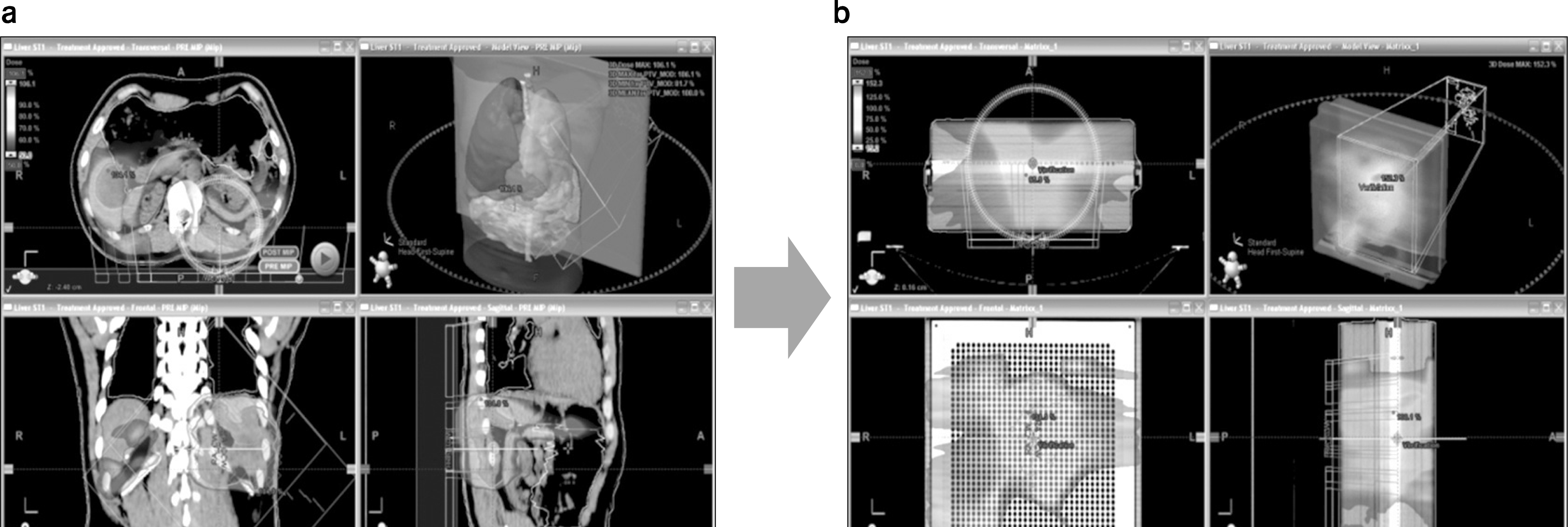
Fig. 2.
(a) Radiation Therapy Planning (RTP) result using by ECLIPSE, (b) Dose distribution of same patient radiation therapy plan when it is projected into MatriXX detector.

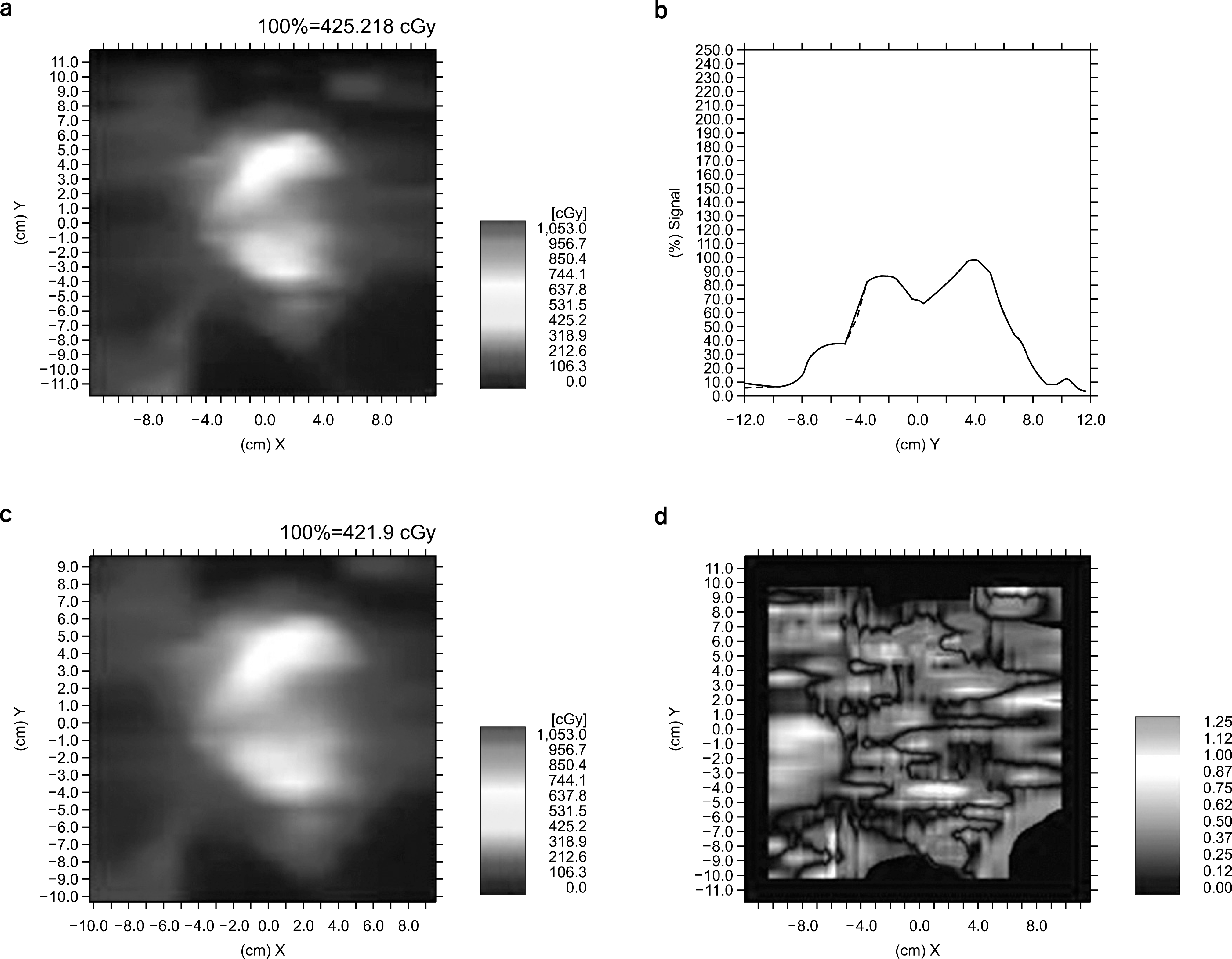
Fig. 3.
(a) 2 dimensional dose distribution of the measurement by MatriXX, (b) dose comparison of x-axis profile between measurement and RTP plan, (c) 2 dimensional dose distribution of the RTP plan by ECLIPSE, (d) gamma distribution for same sample.

Table 1.
Information of the patient radiotherapy plan (1 arc=360o, 2 arc=720o).


 PDF
PDF ePub
ePub Citation
 Print
Print


 XML Download