Abstract
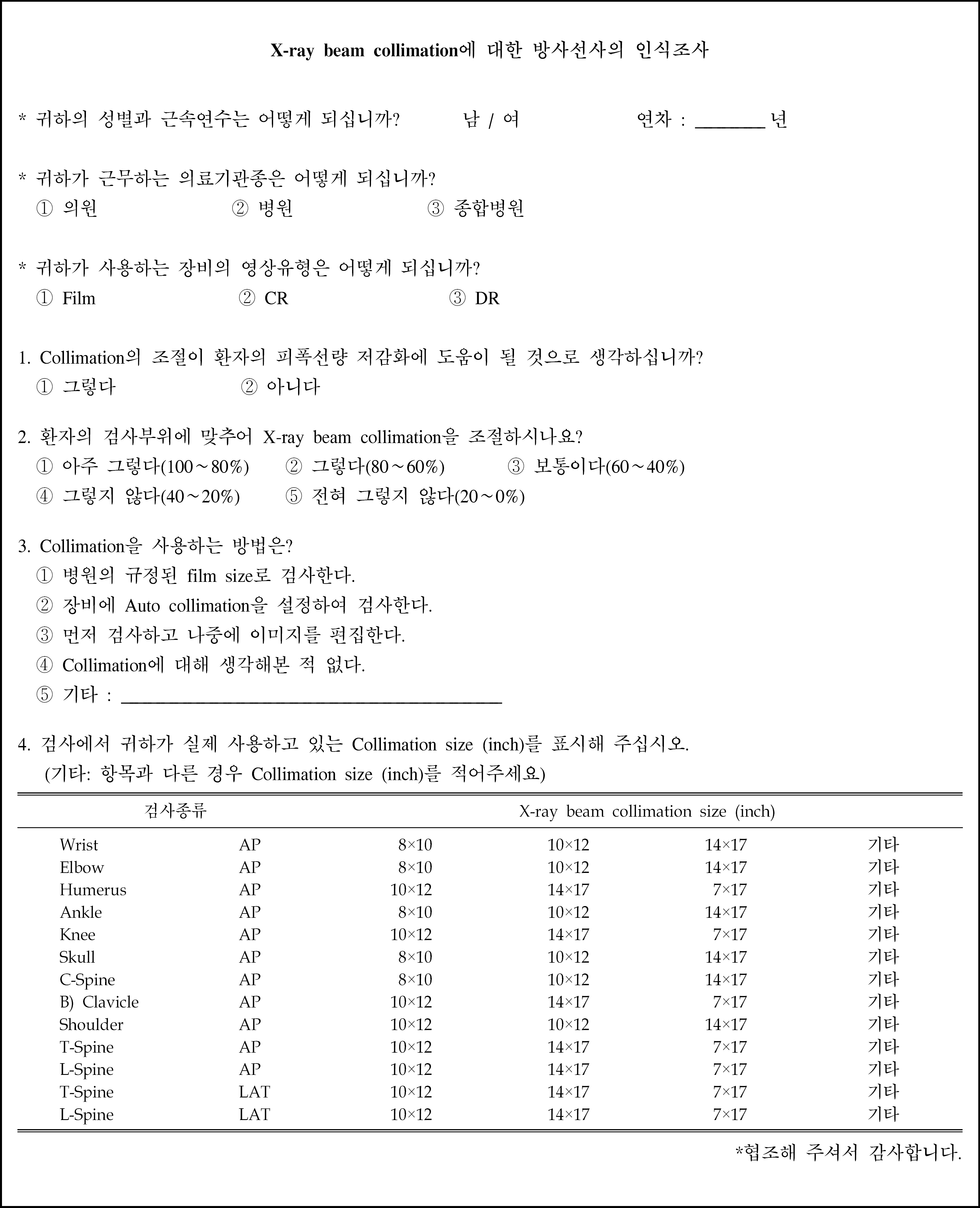
Due to the introduction of CR and DR, it has been neglected the use of the X-ray beam collimator and field size. This study examines nationwide survey of the proper use of collimator and field size by area in a specific field of plain radiography and the current status. Authors emphasized the need for the field size criteria, and propose a standard reference field size in each specific radiologic examination. Total 333 medical institutions (included in Seoul, Gyeonggi-do, Jeolla, Chungcheong, Gangwon-do, Busan area), were investigated in relation to the status of the X-ray beam collimation field size, type specific inspection areas, medical facilities, and image analyses by type to figure out whether they use the adjustment of image field to the specific examination. To assess the awareness and the impact of radiation exposure to the collimation adjustable, 168 radiographers who was working in 10 general hospitals, 10 hospitals, and 10 clinics, were surveyed how they haver adjusted the actual field size. We examine that 61.3% of medical institutions used the “Proper collimation” and only 49.9% of them employed proper one in lumbar spine densely crowded by major organs. 69% among general hospitals, and 65% among hospitals using DR system were using proper collimation. Radiographers recognized that proper adjustment of collimation could reduce the harmful radiation dose on patients. In the survey, 97.6% of respondents were aware of this fact, but only 83.3% of respondents did the adjustment of the size of the collimation field. The using of proper collimation field was low in the nationwide survey, so the effort to reduce the radiation dose on the patients is urgently needed. A unified standard for the field accompanied by thorough education should be needed.
REFERENCES
1. Zetterberg LG, Espeland A. Lumbar spine rediographypoor collimation practices after implementation of digitial technology. Br J Radiol. 84(1002):566–569. 2011.
2. Yoo MK, Kim MS, Lee KJ, Lee J, Beak JM. The optimization method and absorbed dose of L-spine AP by collimation. In Seoul Radiological Technologists Association 118-119. 2009.
3. 임인철, 이상훈. 진단용 X선 발생장치의 X선관 가변조리개 성 능검사와 조사야일치검사 및 중심선속 일치검사에 대한 평가. 한국컨텐츠학회논문지. 9(3):250–254. 2009.
4. Parry RA, Glaze SA, Archer BR. Typical patient radiation doses in diagnostic radiology. Radiographics. 19:1289–1302. 1999.
6. Johnstom JN, Fauber TL. Essentials of radiographic physics and imaging. Elsevier;St. Louis: 2012. ), pp.p. 79–103.
7. Powys R, Robinson J, Kench PL, Ryan J, Brennan PC. Strict X-ray beam collimation for facial bones examination can increase lens exposure. Br J Radiol. 85:e497–e505. 2012.

8. Jeffery CD. The effect of collimation of the irradiated field on objectively meaasured image contrast. Radiography. 3:165–177. 1997.
9. Falk A, Lindhe JE, Rohlin M, Nilsson M. Effects of collimator size of a dental X-ray unit on image contrast. Dentomaxillofac Rad. 28:261–266. 1999.

10. Journy N, Sinno-Tellier S, Maccia C, et al. Main clinical, therapeutic and technical factors related to patient's maximum skin dose in interventional cardiology procedures. BJR. 85:433–442. 2012.

11. Granger WE, Bednarek DR, Rudin S. Primary beam exposure outside the fluoroscopic field of view. Med Phys. 24:703–707. 1997.

Table 2.
X-ray beam collimation for different body regions (total).
| Adequate collimation | Poor collimation | Total | |
|---|---|---|---|
| Partly using | Full open | ||
| 204 (61.3%) | 43 (12.9%) | 86 (25.8%) | 333 (100%) |
Table 3.
X-ray beam collimation for medical institutions.
Table 4.
X-ray beam collimation for kind of image.
Table 5.
X-ray beam collimation for different body regions
Table 6.
Status of X-ray beam collimation size in clinical examinations.
Table 7.
Difference of inadequate rate between clinical and document survey.
Table 8.
Standard X-ray beam collimation size in radiographic examinations.


 PDF
PDF ePub
ePub Citation
 Print
Print


 XML Download