

 PDF
PDF ePub
ePub Citation
 Print
Print


INTRODUCTION
Evaluation of orthodontic tooth movement is one of the most important processes in clinical orthodontics. The experience of treatment and the knowledge from treatment evaluation can increase the clinical competence of orthodontists and improve the treatment outcomes.
Superimposition of cephalograms has traditionally been the most widely used method to evaluate orthodontic tooth movement. However, cephalograms are two-dimensional, and entail difficulty in tracing the overlapped teeth, magnification errors, and radiation exposure.1 Cone-beam computed tomography (CBCT) provides three-dimensional (3D) evaluation; however, it has a relatively low resolution and the limitation of high radiation exposure. Thus, cephalograms and CBCT images are not appropriate for repetitive evaluation during orthodontic treatment.
A digital model can be used to assess tooth position. Previous studies have reported the accuracy and efficacy of digital models for orthodontic analysis.234 Some investigators tried to evaluate orthodontic tooth movement by superimposing digital models obtained from dental casts.5678 However, their evaluations compared only the differences between the pretreatment and posttreatment periods. Consequently, much important information on various tooth movements during treatment, especially during initial leveling and alignment, were overlooked. Periodic and delicate determination of tooth movement is necessary not only to obtain functional and esthetic results, but also to avoid unnecessary tooth movements, such as jiggling movement, and to reduce treatment time, side effects, and costs. To date, no studies have calculated the amount of tooth movement every month during treatment using fixed orthodontic appliances. This may be due to the difficulties in taking impressions without deformation when fixed appliances are attached to the teeth.
Recently, the use of intraoral scanners has enabled the acquisition of digital models by directly scanning the patient's dentition. The purpose of this preliminary study was to test and propose clinical applications of the intraoral scanner for serial evaluation of orthodontic tooth movement.
MATERIALS AND METHODS
Samples
This study was approved by the institutional ethics committee of Gangneung-Wonju National University Dental Hospital (IRB2015-06). The subjects were comprised of eight patients (three males and five females) who were scheduled for comprehensive orthodontic treatment using fixed orthodontic appliances on the labial and/or buccal tooth surfaces. Other inclusion criteria were as follows: no orthodontic appliances and no soft-tissue lesions covering the palate; no missing maxillary central incisor, canine, or first molar; ability to open mouths without difficulty during oral scanning; and agreeing to participate in the study for 4 months. The 0.022-inch edgewise appliances of MBT prescriptions (3M Unitek, Monrovia, CA, USA) were used for orthodontic treatment. Six patients were treated with premolar extraction, and the other two were treated without extraction.
Intraoral scanning
The maxillary dentitions were scanned every month during the initial 4 months of orthodontic treatment: at the beginning of treatment (T0), and at 1 month (T1), 2 months (T2), 3 months (T3), and 4 months (T4) after T0. Intraoral scans were performed using a confocal-type intraoral scanner Trios (3Shape; Copenhagen, Denmark; accuracy: ±7–8 µm) according to the manufacturer's instructions. Briefly, the patients were asked to lean on the dental chair inclined at 45°. Their teeth were dried with gentle air, and the cheeks were retracted for scanning and moisture control. The dentitions were continuously scanned from one side of the posterior teeth to the opposite side along the dentition: initially the occlusal surface, then the buccal and lingual surfaces, and finally the palatal area. The scanning procedure took approximately 10 minutes per dentition.
3D coordinate system and reference points
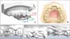
The evaluations of tooth movements in the digital models were performed using Rapidform XOR3 (INUS Technology, Seoul, Korea). A 3D coordinate system with its origin at the incisive papilla was constructed on the T0 digital model (Figure 1A). To transfer the coordinate system of the T0 model to the T1, T2, T3, and T4 models, the digital models were superimposed on the palatal surface (Figure 1B).56 Points 1, 2, and 3 were marked on the base of the brackets or tubes of the bilateral maxillary central incisors, canines, and first molars in each model (Figure 1C–1E). The lines connecting Point 1 and Point 2 were projected to the sagittal plane (X–Y plane) and coronal plane (Y–Z plane) to evaluate the changes in mesiodistal and buccolingual or labiolingual angulation of the central incisor and first molar. The line of the canine was projected to the oblique planes that were rotated 53° from both the sagittal and coronal planes to evaluate the angular changes. The lines connecting Point 2 and Point 3 were projected to the horizontal plane (X–Z plane) for evaluating the rotational changes. The distances from Point 4, midpoint between Point 1 and Point 3, to the sagittal, coronal, and horizontal planes were calculated to evaluate the linear changes of the tooth.
Method error
The maxillary dentitions of seven patients were scanned twice at T0. Thereafter, the reference points were marked and superimpositions were performed to assess the linear and angular differences of the teeth, which should be minimal. The Dahlberg formula was used to calculate the mean error, and the intraclass correlation coefficient (ICC) was calculated to test the repeatability of this method.
RESULTS
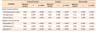
Method errors ranged from 0.04 mm to 0.16 mm for linear displacement and from 0.48° to 0.99° for angulation (Table 1). ICC showed very high correlation (r > 0.978), implying good repeatability.
Table 2 summarizes the tooth movements of eight patients during the initial 4 months of orthodontic treatment. The pattern and amount of tooth movement was not constant and showed large standard deviation. Figure 2 shows the changes of dentition in patients with and without premolar extraction throughout the 4 months. Briefly, in Patient #1, the right first molar was derotated by 2.91°, 1.79°, and 5.72° during T1–T2, T2–T3, and T3–T4, respectively, by using the Goshgarian-type transpalatal arch. The canines were moved and tipped distally by using a 0.012-inch nickel-titanum arch wire, but no canine retraction method was applied. In Patient #2, open coils were inserted between the central incisors and canines for making space for the lateral incisors. The right central incisor moved forward (2.29 mm) and proclined (10.03°) progressively during T0–T3, and then moved backward (0.20 mm) and retroclined (5.79°) during T3–T4 after starting the traction of the lateral incisors at T3.
DISCUSSION
Most previous studies on orthodontic tooth movement have compared only the differences between the pretreatment and posttreatment periods.5678 During treatment, however, clinicians frequently have to evaluate tooth movement to decide whether to change the arch wires and/or biomechanics. However, acquiring repetitive cephalograms and CBCT images increases the risk of radiation exposure. Obtaining a digital model from the dental cast is impractical, because taking an impression, pouring, and scanning every month require much effort, time, and cost. Furthermore, complicated orthodontic appliances may cause distortion and tearing when the impression is removed from the mouth. In contrast, the intraoral scanner directly obtains the digital model from the patient's dentition. Therefore, this contactless scanning may become the solution for patients with fixed orthodontic appliances.
Accuracy and validity of the digital model from the intraoral scanner has already been confirmed in previous studies.910 The present study tested the repeatability of measuring the changes in tooth position by using the reference points on the bracket base in the digital model (Table 1). Mean errors less than 0.16 mm and 0.99° suggest that this method is clinically acceptable. We used the reference points on the bracket or tube base, because the facial axis of the clinical crown (FACC) and the FACC point were not reliable for identification. Therefore, the mesiodistal and labiolingual angles from Point 1 and Point 2 do not indicate the real crown angulation and inclination by FACC, and it should be used only for assessing the changes. Specially constructed jigs allowing brackets to be placed at the same position are recommended, because brackets may be detached during treatment. Pre-marking the reference points on the bracket base will be helpful for identification and improving reliability.
Table 2 shows an inconstant tooth movement and a large standard deviation. The reason may be that the malocclusion of patients was diverse and proper treatment methods were inevitably inconsistent. Moreover, the sample size of the present study was considered somewhat small, even though the minimal sample size was 5 for ICC = 0.95 with significance level (alpha) = 0.05 and width of confidence interval = 0.2.11 Further studies with well-controlled samples and sufficient sample size are needed to determine the amount and pattern of tooth movement in certain malocclusions, when using various appliances and treatment methods.
The present study focused on the clinical application of the intraoral scanner for the serial evaluation of orthodontic tooth movement in patients with fixed appliances. Clinicians cannot easily notice subtle movements during treatment. However, the recognition of such tooth movements may be crucial in improving the treatment quality, and intraoral scanning may be the solution.


 XML Download