

 PDF
PDF ePub
ePub Citation
 Print
Print


INTRODUCTION
The capitate is the largest and most central bone of all the carpal bones (1). Due to its anatomical location, it is well-protected such that capitate fracture rarely occurs, accounting for only 1–3% of all carpal fractures (2). Most capitate fractures are associated with other carpal injuries, such as scaphoid fractures and perilunate injuries. Isolated capitate fractures are very uncommon and are often non-displaced, because of the stabilization by intracarpal ligaments (3). Capitate fractures occur through a number of mechanisms. For instance, falling on one's palm with the wrist extended and getting in a motor vehicle crash are the most commonly cited (3). On the other hand, stress fractures result from either a repeated force by new exercise or by trauma to bone (4).
Although stress fractures of tarsal bones are now frequently diagnosed, their occurrence in the carpal bones are seen far less often because they may be underdiagnosed. The distribution of carpal stress fractures in childhood has been previously reported, among which the capitate is most frequently affected (4). Skeletally immature individuals are more susceptible to stress injury due to weak chondro-osseous junctions, increased physical activity, less muscle mass, and narrower bones with thinner cortices (5). In the case of adults, Vizkelety and Wouters (6) reported the first case of stress fracture of capitate in 1972, and another case was reported in 1994 to date in the literature to the best of our knowledge (7). This report states the radiologic findings of a stress fracture of the capitate of an honor guard and reviews the carpal kinematics related to the stress fracture of the capitate.
CASE REPORT
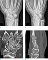
A 20-year-old male reported pain in his right wrist which had continued for five months. The patient reported no traumatic episodes nor any other significant medical or surgical history. However, given the nature of his work serving as an honor guard in the military, he had sustained excessive movement of both wrists (Fig. 1).
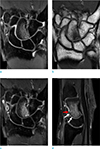
Conventional radiographs and three-dimensional computed tomography (CT) of the right wrist have revealed a minimally displaced fracture line located at the midcarpal aspect of the right capitate (Fig. 2). A magnetic resonance imaging (MRI) scan of the patient has demonstrated a subarticular capitate fracture in the midcarpal aspect with depression and diffuse bone marrow edema (Fig. 3). In addition, small osteophytes were detected in the capitate with irregularity of the midcarpal articular cartilage and a suspicious volar capito-hamate ligament injury.
Regarding the aforementioned findings, we conclude that the patient suffered a stress fracture of the capitate with osteoarthritis of the midcarpal joint and an adjacent ligament injury. Hence, the patient underwent an arthroscopic debridement and multiple drillings using a drill and K-wire. The capitate subchondral fracture and synovial hypertrophy were confirmed during surgery. There were no complications, and the patient was able to resume his daily routine two months post-operation.
DISCUSSION
A review of the literature shows that isolated fracture of the capitate is rare and frequently associated with trauma, most commonly resulting from a fall on the palm with the wrist extended, axial load, or a direct blow over the dorsum of the wrist. The patient did not report any history of acute injury and his symptoms had developed gradually, starting with his military service as an honor guard. These features suggest that the stress fracture was caused by a repetitive micro-trauma rather than a single traumatic event. The case presented in this report, to our knowledge, is the third known case of capitate stress fracture in adults (67).
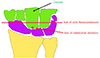
The capitate is often considered as the “keystone” of the carpus, not simply because of its central and prominent position in the wrist, but also because of its mechanical interactions with neighboring bones (8). Previous studies on wrist motion have provided information regarding the functional and mechanical significance of the capitate. According to the findings of these studies, the wrist has two degrees of freedom of motion - flexion-extension motion and radioulnar deviation - with two axes of rotation located within the head of the capitate where one is slightly more proximal than the other (Fig. 4). In a recent study, a rotational motion around the axis has been shown to obliquely penetrate the head of the capitate in an almost radial extension/ulnoflexion plane of motion of the wrist (9). The patient in our case had conducted repeated rotation and flexion/extension of both wrists during his military service as an honor guard.
Stress fractures occur when a normal bone is injured by an abnormal activity (4). The most common cause of stress fracture is a repetition of certain activities. Due to strenuous training activities, military recruits and professional athletes have mostly been the subjects of articles on stress fractures (10). When a bone suffers stress, it responds initially with accelerated cortical resorption and remodeling of the Haversian systems, which as a result weakens the cortex (5). A persisting stress leads to the osteoclastic resorption outpacing osteoblastic repair, which further weakens the cortex and causes fracture unless the stress factor is removed. Stress fracture of carpal bones is rare, among which the capitate is the most frequently affected, followed by the lunate and the scaphoid (4).
In the previous report, stress fracture of the capitate was seen as an irregular fracture line at distal one third of capitate bone, without evidence of avascular necrosis (7). In our case, the fracture line was located at the proximal portion of scaphoid bone and the displacement was less prominent. To our knowledge, specific imaging findings of capitate stress fractures have not yet been reported. In general, MRI is considered the best diagnostic modality for the identification of stress fractures (510). Marrow edema results in low signal intensity on T1-weighted and high signal intensity on STIR (short tau inversion recovery image) and T2-weighted MRI. Sub-periosteal fluid or periosteal edema may be meaningful and ancillary findings in the cases of subtle fractures. In more advanced phases, a fracture line could be well defined by linear low signal intensity on T1- and T2-weighted images. When intravenous contrast agents are administered, the medullary space shows enhancement secondary to edema. In some cases, CT scans may also be effective in conducting a more precise diagnosis of stress fractures, especially when additional information is required for planning surgery. Conventional radiographs are relatively insensitive, demonstrating a 15–35% sensitivity in the initial examination, which increases to 30–70% during follow-up due to a more overt bone reaction.
Although stress fracture of the capitate is rare in adults, we suggest that it should be considered as a differential diagnosis for patients who have conducted repeated motion of the wrist to a considerable extent.

 XML Download