

 PDF
PDF ePub
ePub Citation
 Print
Print


Brachial plexus block can reduce the complications caused by side effects such as difficulty in airway management, retching and vomiting. It also has convenient advantages including postoperative pain management. The technique has thus been performed frequently for surgeries on the upper extremities.
The approach may differ depending on the surgical site. Specific examples include interscalene block, supraclavicular block, subclavicular, axillary block, etc.
The supraclavicular block is widely used as anesthesia for surgeries of the elbow, lower arm, and hand areas because it ensures a precise and quick nerve block even with a relatively low dose of local anesthetic.1
However, complications may occur. Approximately 0.5% to 6% cases of pneumothorax have been reported. Phrenic nerve block (40%–60%) and Horner's Syndrome, neuropathy, etc. may also occur.
Horner's Syndrome is a symptom caused by an abnormality of the sympathetic nervous pathway (oculosympathetic pathway) distributed at and around the eyes. Its clinical features include eyelid ptosis, stenocoriasis, and facial hyphidrosis, and may be classified as central, preganglionic or postganglionic Horner's Syndrome according to the above position.2
The author experienced Horner's Syndrome, which occurred after the brachial plexus block using the supraclavicular block was performed. The author reports the experience with consideration through literature.
CASE
The patient is a 45-year-old woman who is 162 cm tall and weighs 59 kg. About 3 weeks ago, she was diagnosed with Lt. hand 4th Metacarpophalangeal Fx. after she slipped and struck her hand. The patient was admitted to the orthopedic surgery ward to undergo corrective osteotomy and pinning.
There were no exceptional cases of health conditions or disease in her medical history. The preoperative ECG, chest radiography, and blood test were performed, and the results were all normal.
The patient underwent the ultrasound-guided supraclavicular block while being monitored for vital signs. She received a series of 2 injections, 1% lidocaine (11 ml in total) and 0.75% ropivacaine (11 ml in total). The measured doses were administered in the following order: 1% lidocaine (3 ml), 0.75% ropivacaine (3 ml), 1% lidocaine (4 ml), 0.75% ropivacaine (4 ml), 1% lidocaine (4 ml), and 0.75% ropivacaine (4 ml).
As the supraclavicular block was performed, the patient's vital signs were maintained under highly stable conditions. They were recorded as 110/95 mmHg for blood pressure, 36.7℃ for body temperature, 65 BPM, and 14 BRA (Fig. 1).
She complained of difficulty in opening her left eye 20 minutes after the supraclavicular block was performed. Moreover, miosis was observed in the pupil of the left eye.
However, the normal pupil light reflex was maintained in both eyes.
The patient's level of consciousness was clear and vital signs were also normal.
The patient underwent surgery upon being diagnosed with left unilateral Horner's Syndrome, and was monitored for symptoms over time.
The surgery was completed one hour and 50 minutes after the initiation of the supraclavicular block with no reported complications.
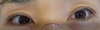
The pupils of both eyes were observed in the recovery room two hours after the procedure, and showed the same size (isocoric) and normal shape (round) (Fig. 2).
There was no bilateral ptosis, and the eyeball moved freely in all directions without any restriction.
The patient eventually recovered from her previous difficulty in closing the left eye, and she did not complain of any discomfort.
She was moved to a general ward after being observed for 30 minutes with no further complications in the recovery room.
DISCUSSION
Local anesthesia minimizes airway manipulation as well as hemodynamic changes associated with general anesthesia. Its advantages include better pain control post-surgery, quick initiation of rehabilitation, decreased stress response to surgery, reduced retching and vomiting following surgery, etc.345
In particular, the brachial plexus block has many advantages in the surgery of the upper extremity due to various advantages of local anesthesia.6
Theoretically, if a local anesthetic is injected into the neurovascular sheath surrounding the brachial plexus, its distribution may block the nerves of the brachial plexus but the actual effects may vary.789
Horner's Syndrome may occur even with the interception of the sympathetic nervous pathway from the hypothalamus to the eyeball.
The sympathetic nervous pathway of the eyes runs from the posterolateral part of the hypothalamus through the brain stem reticular formation, side by side with the lateral spinothalamic tract, and finally ends at the ciliospinal center of Budge-Waller of the intermediolateral gray substance between C8 and T2.
Preganglionic neurons pass through the central nervous system via the nerve roots of the ventral branch, and connect to the postganglionic neuron at the cervical ganglion. The axon of the postganglionic neuron is distributed around the face including the eye socket.
Reported symptoms of Horner's Syndrome symptoms include eyelid ptosis, pupil miosis, and hypohidrosis. Pupil miosis causes a difference in size between the normal and affected pupils, and the difference in pupil size between the eyes decreases when exposed to the light. In addition, the affected pupil shows passive mydriasis due to the relaxation of the sphincter of pupil in the dark, and mydriasis appears more slowly than in the normal pupil.
Hypohidrosis may occur in affected facial areas in the case of preganglionic lesions. While its occurrence is rare in such a case, the facial area that is primarily impacted is above the eyebrows.
In most cases, Horner's Syndrome is diagnosed based on observations of patients' clinical symptoms as well as through a drug test on the pupil. That is, the presence of Horner's Syndrome can be confirmed by checking the sympathetic nerve for damage by cocaine instillation.
Hydroxyamphetamine secretes norepinephrine at the nerve junction to enlarge the pupil. Therefore, if the postganglionic neuron is normal, mydriasis appears due to the presence of hydroxyamphetamine. If the postganglionic neuron is abnormal, it can be determined that damage has been sustained to the central neuron or the preganglionic neuron.10
Since it has not been clinically applied, there are many limitations in carrying out such a test.
In this case, a drug test was not conducted. However, 20 minutes after the supraclavicular block was performed, the patient complained of difficulty in opening her left eye, and miosis was observed in the left pupil. Based only on these symptoms, it was reasonable to diagnose the patient with left unilateral Horner's Syndrome.
Horner's Syndrome has been reported to occur following thoracostomy, internal jugular vein cannulation, Swan-Ganz catheterization, and brachial plexus block using axillary block.11121314
In conclusion, a 45-year-old female patient showed symptoms distinctively attributed to Horner's Syndrome. She complained of ipsilateral ptosis and pupil miosis 20 minutes after receiving the supraclavicular block, but her condition was immediately restored two hours later. Although the exact mechanism is unknown, the author considers that the cause lies not in the dose of the local anesthetic. Rather, it may be in relation to the method in which the local anesthetic was employed. The needle used to inject the local anesthetic was positioned around the center of the vertebrae past the inner side of the long platysma. The latter is believed to have caused distinct Horner's Syndrome in the ipsilateral pupil.15
Although the supraclavicular block is a relatively simple procedure that can be performed easily, it may cause several side effects. Therefore, it is necessary to make thorough preparations prior to administration and fully inform patients of potential side effects.
Medical professionals should respond quickly in the event that pupil miosis or eyelid ptosis occurs following the supraclavicular block. They are advised to check for the appearance of Horner's Syndrome as quickly as possible to reduce patient anxiety and discomfort.

 XML Download