

 PDF
PDF ePub
ePub Citation
 Print
Print


Abstract
Objectives
To determine the relationship between lumbar disc degeneration and back muscle degeneration.
Summary of Literature Review
In the degenerative cascade of the spine described by Kirkaldy-Willis, degeneration of the disc and of the facet joint co-occur with aging. However, the muscles of the back are not included in this model. Several studies have reported significant correlations between back muscle degeneration and facet joint arthritis. The purpose of our study was to evaluate relationships between lumbar disc degeneration and fatty degeneration of the back muscles.
Materials and Methods
In this study, 65 patients over the age of 50 years who had undergone lumbar spine MRI in our orthopaedic clinic were recruited. Fatty degeneration of the back muscles was qualitatively graded from I to III by the degree of the fat signal in the muscle layer, including both the multifidus and erector spinae. Lumbar disc degeneration was graded from I to V according to the Pfirrmann grade. Correlations between the back muscle degeneration grade and radiological parameters were analyzed.
Results
The degeneration grade of the multifidus correlated positively with age and the grade of disc degeneration. Correlations with other radiologic parameters were not significant. The degeneration grade of the erector spinae correlated positively with age. Other radiologic parameters were not significant.
Conclusions
There was a significant correlation between lumbar disc degeneration and multifidus degeneration. Erector spinae degeneration was correlated with age, but not with lumbar disc degeneration. The degenerative cascade of the spine was accompanied by fatty changes of the multifidus with aging.
REFERENCES
1. Kirkaldy-Willis WH, Wedge JH, Yong-Hing K, et al. Pathology and pathogenesis of lumbar spondylosis and stenosis. Spine (Phila Pa 1976). 1978 Dec; 3(4):319–28. DOI: DOI:10.1097/00007632-197812000-00004.

2. Kalichman L, Klindukhov A, Li L, et al. Indices of Paraspinal Muscles Degeneration: Reliability and Association With Facet Joint Osteoarthritis: Feasibility Study. Clin Spine Surg. 2016 Nov; 29(9):465–70. DOI: 10.1097/BSD.0b013e31828be943.
3. Tsekoura M, Kastrinis A, Katsoulaki M, et al. Sarcope-nia and Its Impact on Quality of Life. Adv Exp Med Biol. 2017; 987:213–8. DOI: 10.1007/978-3-319-57379-3_19.
4. Shahidi B, Parra CL, Berry DB, et al. Contribution of Lumbar Spine Pathology and age to Paraspinal Muscle Size and fatty Infiltration. Spine (Phila Pa 1976). 2017 Apr 15; 42(8):616–23. DOI: 10.1097/BRS.0000000000001848.
5. Legaye J, Duval-Beaupere G, Hecquet J, et al. Pelvic incidence: a fundamental pelvic parameter for three-dimensional regulation of spinal sagittal curves. Eur Spine J. 1998 May; 7(2):99–103. DOI: DOI:10.1007/s005860050038.
6. Pfirrmann CW, Metzdorf A, Zanetti M, et al. Magnetic resonance classification of lumbar intervertebral disc degeneration. Spine (Phila Pa 1976). 2001 Sep 1; 26(17):1873–8. DOI: DOI:10.1097/00007632-200109010-00011.
7. Lee JC, Cha JG, Kim Y, et al. Quantitative analysis of back muscle degeneration in the patients with the degenerative lumbar flat back using a digital image analysis: comparison with the normal controls. Spine (Phila Pa 1976). 2008 Feb 1; 33(3):318–25. DOI: 10.1097/BRS.0b013e318162458f.
8. Kjaer P, Bendix T, Sorensen JS, et al. Are MRI-defined fat infiltrations in the multifidus muscles associated with low back pain? BMC Med. 2007 Jan 25; 5(1):2. DOI: 10.1186/1741-7015-5-2.
9. Landis JR, Koch GG. The measurement of observer agreement for categorical data. Biometrics. 1977 Mar; 33(1):15974. DOI: DOI:10.2307/2529310.
10. Freeman MD, Woodham MA, Woodham AW. The role of the lumbar multifidus in chronic low back pain: a review. PM R. 2010 Feb; 2(2):142–6. DOI: 10.1016/j.pmrj.2009.11.006.
11. Mannion AF. Fibre type characteristics and function of the human paraspinal muscles: normal values and changes in association with low back pain. J Electromyogr Ki-nesiol. 1999 Dec; 9(6):363–77. DOI: DOI:10.1016/s1050-6411 (99)00010-3.
12. Bogduk N. A reappraisal of the anatomy of the human lumbar erector spinae. J Anat. 1980 Oct; 131(Pt 3):525–40.
13. Ohtori S, Orita S, Yamauchi K, et al. Classification of Chronic Back Muscle Degeneration after Spinal Surgery and Its Relationship with Low Back Pain. Asian Spine J. 2016 Jun; 10(3):516–21. DOI: 10.4184/asj.2016.10.3.516.
14. Crawford RJ, Filli L, Elliott JM, et al. Age- and Level-Dependence of Fatty Infiltration in Lumbar Paravertebral Muscles of Healthy Volunteers. AJNR Am J Neuroradiol. 2016 Apr; 37(4):742–8. DOI: 10.3174/ajnr.A4596.
Fig. 1.
Qualitative analysis of the fatty infiltration of lumbar back muscles. (A) Grade 1: mild (<10% fatty infiltration). (B) Grade 2: moderate (10-50% fatty infiltration). (C) Grade 3: severe (>50% fatty infiltration). Black arrow: multifidus, dotted arrow: erector spinae.
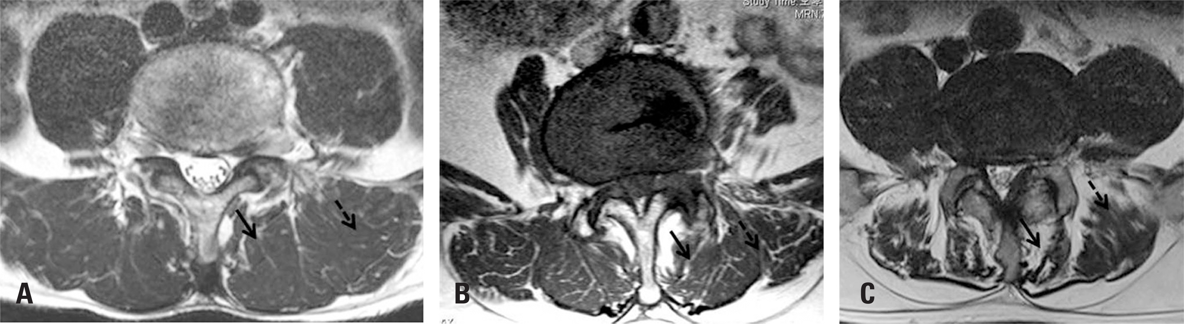
Table 1.
Descriptive data of patients
Table 2.
Correlation analysis between radiological factor and fat infiltration(FI) grade of multifidus
Table 3.
Correlation analysis between radiological factor and fat infiltration(FI) grade of erector spinae
 XML Download