

 PDF
PDF ePub
ePub Citation
 Print
Print


INTRODUCTION
Crohn disease (CD) is characterized by inflammatory mucosal lesions that affect all regions of the gastrointestinal (GI) tract [1]. Primary perianal lesions are caused by bowel inflammation, and include superficial rupture, anal ulcers, and lymphedema [23]. Fistulas and abscesses are primary lesions. Anal stenosis is associated with primary or secondary perianal lesions, and is a long-term consequence of inflammation [2]. Perianal CD can be classified as fistulizing or non-fistulizing [4]. Fistula and abscesses are commonly reported among adults with perianal CD [56]. Reports on pediatric perianal CD are relatively rare, and are mostly regarding the clinical progress and treatment of perianal lesions [27].
The incidence of perianal fistula varies depending on the location of CD, but can increase by up to 92% if colorectal lesions are included in the assessment of occurrence [8]. In addition, perianal fistulas can be detected as the first sign of CD before other symptoms develop. Indeed, perianal fistulas can form years before CD symptoms or pathological changes occur [910].
A number of studies have reported high incidence rates of anal stenosis and anal ulcers among patients with CD [611]. In a study including children and adolescents, 41 of 276 patients diagnosed with CD showed perianal lesions within 30 days after diagnosis. These patients had abscesses, fissures, and skin tags in addition to fistulas [27].
In Korea, the incidence of pediatric CD is gradually increasing [12]. However, there is scarcity of data on perianal lesions in Korean children with CD. Thus, in this study, we aimed to investigate the characteristics of pediatric CD with accompanying perianal lesions.
MATERIALS AND METHODS
Subjects and methods
The study protocol was approved by the Institutional Review Boards of Gachon University Gil Medical Center (IRB number GAIRB2017-237).
Of the 76 patients diagnosed with CD at Gachon University Gil Medical Center between January 2000 and December 2014, 5 patients without available GI endoscopic findings, and 2 patients whose diagnoses werelater changed to ulcerative colitis were excluded. The remaining 69 pediatric and adolescent patients (age ≤18 years) were retrospectively analyzed.
We reviewed the medical records of the patients including, abdominopelvic computed tomography images, colonoscopic findings, wireless capsule endoscopic findings, and histopathology. The following demographic and clinical information was collected: sex, height, weight, chief complaint, presence of associated symptoms, and presence of perianal lesions. Laboratory findings, such ascomplete blood cell count, erythrocyte sedimentation rate, C-reactive protein, and albumin, were extracted. We used physical exams and colonoscopy to confirm perianal lesions.The location and behavior of CD were evaluated using the Montreal classification. Perianal perforating lesions included fistula and abscesses [56]. We also included skin tags along with fistulas and abscesses as perianal lesions [234712]. Comparison of clinicodemographic characteristics between groups was conducted in two separate analyses. In the first analysis, patients were divided into two groups according to the presence or absence of perianal lesions. In the second analysis, they were divided according to the presence or absence of perianal perforating lesions.
Statistical analysis
Either the chi-squared test or Fisher's exact test was used for categorical variables, and the Mann-Whitney U-test or Student's t-test were used to compare continuous variables. All statistical analyses and calculations were performed using IBM SPSS Statistics for Windows ver. 21.0 (IBM Co., Armonk, NY, USA). The level of statistical significance was set at p<0.05.
RESULTS
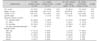
Among the 69 patients with CD (mean age, 15.4 years), 51 (73.9%) were male, 54 had CD with perianal lesions (78.3%) and 29 had perianal perforating lesions (42.0%). No significant differences in the demographic characteristics were found between the two groups. No significant differences in location were found between the groups having CD with and without perianal lesions, as well as between the groups having CD with and without perianal perforating lesions (Table 1).
Abdominal pain was the most common chief complaint at the time of diagnosis. Other symptoms included perianal pain, anal discharge, weight loss, and general weakness. The median symptom duration in CD patients with perianal lesion was longer than in patients without perianal lesions. However, no significant differences in median symptom duration were found between CD with and without perianal perforating lesions. All patients had abdominal symptoms upon diagnosis. Perianal symptoms were observed to preceded GI symptoms in 13 of 29 (44.8%) patients with perianal perforating lesions and the median duration was 14 months (Table 2).
DISCUSSION
We did not find any significant factors of pediatric CD with perianal lesion, but found that perianal symptoms preceded abdominal symptoms in approximately half of the children with perianal perforating lesions and that median duration of chief complaints was longer in pediatric CD patients with accompanying perianal lesions.
In previous studies, perianal lesions of CD included only perianal fistula and abscess [568910]. However, we also considered skin tags along with fistulas and abscesses to be perianal lesions [234712]. The proportion of CD patients with perianal lesions, including skin tag, was high compared with incidence rates reported in other studies [2712].
The most common initial symptoms of CD are nonspecific: chronic abdominal pain, diarrhea, and failure to gain weight [131415]. Abdominal pain in particular is a typical complaint among children and adolescents [16]. However, perianal fistulas can be detected as the first sign of CD before other symptoms and may form years before CD symptoms or pathological changes occur, as reported in other studies [910]. Forty-five percent of the patients developed a perianal fistula before, or at the time of, the diagnosis of CD in a study of CD in Western adults [10]. In a study of CD patients in Korean adults, perianal fistulas were present before or at diagnosis of CD in 36.7% of patients, and were the initial presentation of CD in 15.8% of patients [17]. We also found that there were many cases in which perianal symptoms predicted GI symptom development. Perianal symptoms that precede typical GI symptoms may be an interesting topic for future investigations.
A demographic study reported a mean time of 12 months from symptom onset to diagnosis of in Korean adults, but a recent national cohort study reported a mean time of 5 months [1819]. This trend could easily be observed in a study that classified cohorts of patients with CD according to the year of their diagnosis. The mean time until diagnosis was significantly different between cohorts: 24 months for cohorts diagnosed in 1981–2000 and 14 months for those diagnosed in 2006–2012 [20]. The median time from symptom onset to diagnosis among Korean children was 18 months in 1992, and 7.1 months in 2010 [1121]. In the present study, the median durations were shorter compared with those reported in previous studies [18]. The shorter time from symptom onset to diagnosis may be the result of advanced diagnostic techniques and increased awareness of CD with perianal lesions. In this study, the median duration of chief complaint was longer in children having CD with perianal lesions than those without perianal lesions. However, when patients were divided according to the presence or absence of perianal perforating lesions, no significant difference in symptom duration was observed. Further research is needed to understand the implications of this finding.
The proportion of patients with perianal skin tags in this study was relatively high compared to 21% to 34%, as reported in previous studies [1122]. Therefore, not only careful examination for perianal lesions including skin tags but also suspicion of CD and subsequent evaluation is required. Further investigation is required on whether the incidence of perianal lesions is truly higher in children than adults among patients with CD, as well as the reason for the high incidence.
This study has some limitations. First, a relatively small number of patients were included in this retrospective study. Second, identification of perianal lesions was done only by inspection and colonoscopy. Third, although colonoscopies were conducted in all the patients, the majority were not fully evaluated throughout the entire GI tract. Due to these limitations, the data from this study should be interpreted with caution. Future prospective, large-scale, multi-center research is required to better reveal the characteristics of perianal lesions and its association with other clinicodemographic factors.
In conclusion, perianal symptoms preceded abdominal symptoms in approximately half of the children with perianal perforating lesions. This finding suggests that careful attention is required in children with perianal lesions, regardless of GI symptoms.

 XML Download