Dear Editor,
Rapid-onset dystonia-parkinsonism (RDP) (also classified as DYT12) is caused by ATP1A3 mutations,1 and usually appears during adolescence or early adulthood.23 Although a recent study showed that patients with the RDP phenotype were highly likely to have an ATP1A3 mutation, some cases did not.2 Herein we report an 81-year-old woman who presented with hemidystonia-parkinsonism that progressed rapidly over the course of 1 week. To our knowledge this is the first report of an elderly patient presenting RDP-mimicking features.
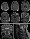
An 81-year-old woman developed involuntary muscle contractions that began in the left fingers and progressed rapidly into the left arm, leg, and neck over the course of 1 week, and then stabilized. Her symptoms appeared after the physical stress associated with helping to hold her husband while he went to the bathroom. Her left eyeball had been removed due to an unknown etiology when she was in her 20s. Although she had severe bilateral knee osteoarthritis, she did not take any medication at the time of symptom onset. Her family—including her parents, siblings, children, and grandchildren—had no history of parkinsonism or dystonia. Two weeks after symptom onset she was admitted to our hospital due to a sustained course of left-side dystonia-parkinsonism that affected the left arm the most severely, followed by the left leg and left neck (first part of Supplementary Video 1 in the online-only Data Supplement). She complained of subjective dysarthria and dysphagia, but these conditions were not clinically significant. The patient had no history of memory impairment or psychiatric disorders including depression. A cognitive assessment using the Seoul Neuropsychological Screening Battery showed no cognitive impairment. Laboratory findings were unremarkable. Brain magnetic resonance imaging (MRI) and whole-spine MRI produced no findings to which the symptoms could be attributed (Fig. 1). Whole-exome sequencing revealed no pathogenic mutation, including in the ATP1A3 gene.
Initially levodopa was prescribed at 300 mg/day, but this was discontinued due to unresponsiveness. We subsequently administered baclofen, procyclidine, and clonazepam, which resulted in a moderate improvement of hemidystonia-parkinsonism on the left side of her body (middle part of Supplementary Video 1 in the online-only Data Supplement). Freezing of gait developed after 2 weeks of treatment. The readministration of levodopa improved this gait freezing, while procyclidine was withdrawn because of memory impairment. Her dystonia-parkinsonism was considerably relieved 2 months later (latter half of Supplementary Video 1 in the online-only Data Supplement) and showed no aggravation during an 18-month follow-up.
Studies indicate that clinical features of RDP with ATP1A3 mutations comprise asymmetry, rapid onset and stabilization within several weeks, predominant dystonia with a rostrocaudal gradient including prominent bulbar symptoms, no tremor, lack of pain, stressful triggers, and no or minimal improvement. Based on these symptoms we considered that our patient presented with rapidly progressing dystonia-parkinsonism, and with stabilization on the left side of her body. Considering both the clinical manifestation and the remarkable improvement, we could rule out synucleinopathy-associated disorders and tauopathy-related disorders including corticobasal syndrome. Further diagnostic brain and whole-spine MRI ruled out secondary etiologies including stroke, tumors, and inflammation. We believe that the symptom presentation combined with MRI findings were most compatible with a diagnosis of RDP among primary dystonia and dystonia-plus syndromes, and so performed whole-exome sequencing to identify any mutation in ATP1A3. However, certain features of this case were notably disparate from classical RDP, with the patient 1) not exhibiting a rostrocaudal gradient, 2) feeling some pain in her left arm and neck, and 3) showing considerable improvement with treatment. In addition, the exact pathomechanism underlying this phenotype remains uncertain, although we deduced that physical stress could have triggered the hemidystonia-parkinsonism in our patient.
The most-striking characteristic of the current case was the age of the patient (81 years). To the best of our knowledge this is the first reported geriatric presentation of the RDP-like phenotype, with the latest age at onset for the RDP phenotype in previous reports being in the 50s.23 Based on the clinical presentation of this case, we considered that there was a low probability of any other known disease being present.4 Moreover, we excluded other known genetic mutations—including for dystonia and parkinsonism—by performing wholeexome sequencing analysis, although this method cannot delineate large deletions of DNA or the existence of trinucleotide-repeat diseases. While the exact etiology of the patient remains unknown, the current findings suggest that elderly patients can present with RDP-mimicking features in the absence of ATP1A3 mutations, and this is clinically relevant to patient diagnosis and care.
Video 1. Initial presentation and serial follow-ups of hemidystonia-parkinsonism. The patient exhibited left-side hemidystonia-parkinsonism, predominantly in the left arm. Left-side hemidystonic gait was observed while walking, although a significant contributing factor to the gait difficulty was severe bilateral osteoarthritis (the first part). Three weeks after treatment the patient exhibited a modest improvement of hemidystonia-parkinsonism (the middle part), and 2 months later the patient showed a remarkable improvement (the latter part).
Figures and Tables
Fig. 1
Brain and cervical MRI of the patient revealed no responsible lesion for hemidystonia-parkinsonism. A and B: Mild cerebral ischemia in periventricular white matter with multifocal leukoaraiosis is evident. However, no hemispheric atrophy or any lesion in the basal ganglia or brainstem is observed. C: Sagittal cervical MRI shows mild cervical disc herniation at the C5 and C6 level, without any cervical myelopathy or root compression. MRI: magnetic resonance imaging.

Supplementary Materials
The online-only Data Supplement is available with this article at https://doi.org/10.3988/jcn.2018.14.3.423.
References
1. de Carvalho Aguiar P, Sweadner KJ, Penniston JT, Zaremba J, Liu L, Caton M, et al. Mutations in the Na+/K+ -ATPase alpha3 gene ATP1A3 are associated with rapid-onset dystonia parkinsonism. Neuron. 2004; 43:169–175.

2. Brashear A, Dobyns WB, de Carvalho Aguiar P, Borg M, Frijns CJ, Gollamudi S, et al. The phenotypic spectrum of rapid-onset dystoniaparkinsonism (RDP) and mutations in the ATP1A3 gene. Brain. 2007; 130:828–835.


 PDF
PDF ePub
ePub Citation
 Print
Print


 XML Download