Journal List > Korean Circ J > v.48(7) > 1097666

Lee, Lee, Lee, Lee, Kang, and Shin: Assessment of Tissue Perfusion with Blood Oxygenation Level-Dependent Magnetic Resonance Imaging in Critical Limb Ischemia
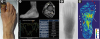
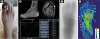
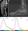
A 62-year-old man with diabetes and end-stage renal disease was referred to our clinic for the management of a nonhealing wound in his right big toe (Figure 1A). Blood oxygenation level-dependent magnetic resonance (BOLD MR) foot images were obtained in conjunction with T2-weighted imaging, time-of-flight magnetic resonance (MR) angiography, and phase-contrast blood velocity mapping using a 3-T MR scanner. These tests revealed absent flow in the anterior tibial artery (ATA) and loss of arterial flow pattern in all infrapopliteal arteries (Figure 1B). Digital subtraction angiography showed diffuse segmental occlusion of all 3 major arteries from the proximal level, with reconstituted flow observed at the distal segment of the ATA (Figure 1C). Successful endovascular revascularization was performed for the ATA with restoration of flow to the lateral tarsal and first dorsal metatarsal arteries (Figure 2C). The skin perfusion pressure value measured near the wound at the dorsal surface increased from 8 to 56 mmHg after the procedure. Compared with the preprocedural one, the postprocedural phase-contrast MR angiography revealed restoration of the arterial flow pattern of the ATA (Figure 2B) and the BOLD MR revealed improved oxygenation (increased T2* values) mainly in the soft tissues of proximal half of the big toe, lateral foot border, and at the intrinsic foot muscles (Figures 1D and 2D). However, the tissue oxygenation failed to improve at the distal half of the big toe (an area of tissue gangrene, Figure 2A). The distribution of individual T2* values assessed from the whole foot (Figure 3C) as well as from the forefoot (Figure 3D) shifted to the right (increasing T2* values) after the procedure.1) The patient underwent first-toe amputation at the basal level of the proximal phalanx, and the amputation stump healed well without any complication.
The signal intensity in BOLD MR images has been shown to correlate well with the oxygenation degree in foot musculature.1) BOLD MR imaging in the foot can allow clinicians to visualize the degree of oxygenation at a glance or directly in the region of interest. It can also help to semiquantify the severity of perfusion deficit by assessing the T2* value.2)3) Our case emphasizes the potential utility of BOLD MR for the volumetric assessment of regional tissue oxygenation in the feet of patients with critical limb ischemia (CLI).4)

Figures and Tables

Figure 1

(A) Clinical photograph shows dry gangrene at the distal half of the toe. (B) Preprocedural time-of-flight magnetic resonance angiography image showing absent anterior tibial artery (ATA) flow. Abnormal arterial waveform was noted in 3 major arteries on phase-contrast blood velocity mapping (yellow line, ATA; orange line, posterior tibial artery [PTA]; green line, peroneal artery [PA]). (C) Digital subtraction angiography image showing no contrast filling along the dorsalis pedis artery. (D) Preprocedural T2* perfusion map of the patient's foot.

kcj-48-658-g001
Figure 2

(A) Clinical photograph after endovascular treatment. (B) Postprocedural time-of-flight magnetic resonance angiography image demonstrating restored anterior tibial artery (ATA) and dorsalis pedis artery flow. The phase-contrast blood velocity map shows improved mean flux and mean velocity of the ATA with improved arterial waveform. (C) After endovascular treatment, flow at the ATA and dorsalis pedis artery was restored. Note the presence of lateral tarsal and dorsal metatarsal arteries. (D) Postprocedural T2* perfusion map. Compared with preprocedural findings, the T2* value increased at the intrinsic foot muscles (long arrows) and soft tissues at the level of the first proximal phalanx and lateral foot border (white arrowheads). Note that the T2* signal intensity in the distal phalangeal area remained low (red arrowheads).

PA = peroneal artery; PTA = posterior tibial artery.
kcj-48-658-g002
Figure 3

(A) T2-weighted axial image used as a reference image for interpretation. (B) The level of the axial image for Figures 1D, 2D, and 3A noted on the sagittal scout image. (C) Histogram of the T2* value of the whole foot, depicted using all T2* values obtained from 15 axial cuts of blood oxygenation level-dependent magnetic resonance images of the foot (slice number 15–29 on Figure 3B). Note that the histogram shifted to the right after endovascular therapy (blue parabola, preprocedural image; red parabola, postprocedural image). (D) Histogram of the T2* value of the forefoot. The forefoot was segmented using the proximal point of the metatarsal bones as a cutoff point.

kcj-48-658-g003

Notes

Funding This work was supported by the Korean Cardiac Research Foundation (201702-01).

Conflict of Interest The authors have no financial conflicts of interest.

Author Contributions

  • Conceptualization: Lee PH, Lee SW, Lee WJ, Shin MJ.

  • Data curation: Lee PH, Lee SW.

  • Formal analysis: Lee JY, Kang JW.

  • Funding acquisition: Lee PH.

  • Investigation: Lee PH, Shin MJ.

  • Methodology: Lee PH, Lee SW, Lee WJ, Kang JW, Shin MJ.

  • Supervision: Lee PH, Lee SW, Shin MJ.

  • Validation: Lee PH, Lee JY, Lee SW, Lee WJ, Kang JW, Shin MJ.

  • Writing - original draft: Lee JY.

  • Writing - review & editing: Lee PH.

References

1. Stacy MR, Qiu M, Papademetris X, Caracciolo CM, Constable RT, Sinusas AJ. Application of BOLD magnetic resonance imaging for evaluating regional volumetric foot tissue oxygenation: a feasibility study in healthy volunteers. Eur J Vasc Endovasc Surg. 2016; 51:743–749.
crossref
2. Bajwa A, Wesolowski R, Patel A, et al. Blood oxygenation level-dependent CMR-derived measures in critical limb ischemia and changes with revascularization. J Am Coll Cardiol. 2016; 67:420–431.
3. Ledermann HP, Heidecker HG, Schulte AC, et al. Calf muscles imaged at BOLD MR: correlation with TcPO2 and flowmetry measurements during ischemia and reactive hyperemia--initial experience. Radiology. 2006; 241:477–484.
4. Bajwa A, Wesolowski R, Patel A, et al. Assessment of tissue perfusion in the lower limb: current methods and techniques under development. Circ Cardiovasc Imaging. 2014; 7:836–843.
TOOLS
ORCID iDs

Joo Yeon Lee
https://orcid.org/0000-0001-8964-7107

Pil Hyung Lee
https://orcid.org/0000-0001-7339-9188

Seung-Whan Lee
https://orcid.org/0000-0002-9301-8183

Woo Je Lee
https://orcid.org/0000-0002-9605-9693

Joon-Won Kang
https://orcid.org/0000-0001-6478-0390

Myung Jin Shin
https://orcid.org/0000-0001-8016-2891

Similar articles