

 PDF
PDF ePub
ePub Citation
 Print
Print


INTRODUCTION
Multiple myeloma (MM) is the second most common hematological malignancy (13%), accounting for 1% of all neoplasms, and is characterized by neoplastic proliferation of plasma cells (1). MM is usually confined to the bone marrow, with 80–90% of MM patients exhibiting skeletal involvement. Extramedullary spread is relatively rare; the incidence is only 3–5% (2). Central nervous system involvement in multiple myeloma (CNS-MM) is an uncommon condition developing in 1% of MM patients. CNS-MM is diagnosed by detection of monoclonal plasma cells in the cerebrospinal fluid (CSF) or CNS involvement evident on neuroimaging of previously diagnosed MM patients (3). CNS-MM may manifest as a dural, parenchymal or leptomeningeal lesion (45). However, only a single case of CNS-MM manifesting as an intraventricular mass has been reported in the literature (6).
Herein, we report a case of CNS-MM in a patient with 17p13 deletion (del 17p) manifested as an intraventricular mass with leptomeningeal involvement combined with perineural spread. This is the first reported case of a CNS-MM featuring a contemporaneous intraventricular mass and leptomeningeal involvement; both are very rare manifestations of CNS-MM. We also describe the unusual computed tomography (CT) and magnetic resonance imaging (MRI) findings.
CASE REPORT
A 66-year-old female visited our hospital complaining of dyspnea. The initial laboratory findings revealed anemia (Hb 9.6 g/dL; reference range 12–16) and an abnormal serum albumin/globulin ratio (albumin 3.6 g/dL, globulin 11 g/dL, normal ratio 1:1) leading to further work up. Serum and urine protein electrophoresis were performed, and serum M-spike was detected (5.4 g/dL). Serum and urine immunofixation electrophoresis revealed IgG λ-type monoclonal gammopathy. The serum beta-2 microglobulin and lactate dehydrogenase were 2.9 mg/L (reference range 0–2.4) and 178 U/mL (reference range 116–243), respectively. Bone marrow examination revealed 40.1% plasma cells. On cytogenetic analysis, a complex karyotype including del 17p was detected. Finally, she was diagnosed as IgG λ-type MM with high-risk cytogenetic features.
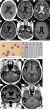
She had undergone chemotherapy, but during the treatment, spine involvement of MM was newly developed leading to palliative radiotherapy on T-spines. Despite the subsequent chemotherapy of higher lines, the disease progressed with extramedullary spread involving soft tissues of the trunk. Twenty months after the initial diagnosis, she presented with headache, nausea, vomiting, and slurred speech. Non-contrast-enhanced brain CT displayed an ovoid hyperdense mass (50 Hounsfield units) in the trigone of the left lateral ventricle (Fig. 1A1), reflecting hypercellular tumor, which was not seen on previous MRI performed 8 months ago (Fig. 1A6). Brain MRI revealed an isointense signal in the mass on both T1- and T2-weighted images with strong homogeneous enhancement (Fig. 1A2–1A4). On susceptibility weighted imaging, it showed a few hemorrhagic foci in the mass. Contrast-enhanced T1-weighted imaging revealed leptomeningeal enhancement along both cerebellar folia (Fig. 1A5). These findings suggested intraventricular and leptomeningeal involvement of the systemic MM, considering her recent medical history. CSF analysis revealed abundant malignant plasma cells (Fig. 1B1) and a white blood cell count of 300/µL (monocytes 5%, neoplastic plasma cells 95%). CSF electrophoresis (Fig. 1B2) confirmed the presence of IgG λ-type M-protein. She received intrathecal chemotherapy and whole-brain radiotherapy to treat the CNS disease, with systemic chemotherapy. Approximately 2 weeks from the commencement of intrathecal chemotherapy, she complained of diplopia, and follow-up MRI was performed. The previously noted intraventricular mass exhibited no significant change; however, leptomeningeal enhancing lesions had progressed along the brain surface of both posterior fossae and the supratentorial region (Fig. 1C1–1C3). Abnormal enhancement of the cisternal segments of both trigeminal nerves with focal extension into the left Meckel's cave was noted, and abnormally enhancing lesions were also noted along the maxillary segments of both trigeminal nerves. Both cavernous sinuses exhibited bulging contours with strong enhancement (Fig. 1C4). These imaging findings suggested aggravated leptomeningeal dissemination and perineural spread of the known CNS-MM. Unfortunately, her condition deteriorated further, and she died 2 years after diagnosis and 2 months after the initial neurological manifestations.
DISCUSSION
CNS-MM is a very rare condition with incidence of 1% in MM patients, and exhibits aggressive terminal disease feature, often associated with poor prognosis (37). This is the first report of a CNS-MM patient with an intraventricular mass and leptomeningeal involvement. Various manifestations of CNS-MM have been reported; these include dural, parenchymal and leptomeningeal involvement. Most of them manifest as dural involvement, most commonly resulting from osseous lesions in skull, while primary dural involvement is rarer (45). Intraventricular involvement of CNS-MM, meanwhile, is far rarer, and only a single case report has been published. Eum et al. (6) reported a case of MM manifesting primarily as a lateral ventricular mass. On imaging, a large, homogeneously contrast-enhancing intraventricular mass with hydrocephalus was evident. In our present case, the CNS-MM also manifested as a strong, homogeneously enhancing intraventricular mass with intratumoral hemorrhagic foci. The signal intensity of the intraventricular tumor was similar to that of dural MM tumors and to that of the only report of MM manifesting as an intraventricular tumor (cited above). However, such a finding is non-specific, and the possibility of a subependymoma, intraventricular metastasis, a meningioma, or a choroid plexus tumor must all be excluded, given the location of the tumor. A CNN-MM diagnosis is usually made when the lesion appears in the context of previously diagnosed MM and is confirmed by CSF analysis or tissue biopsy, stereotactic biopsy or resection. Although we did not perform diffusion-weighted imaging on our patient, diffusion restriction may have been evident, as is true of MM with dural involvement. This is attributable to the high cellularity and low nucleocytoplasmic ratio of CNS-MM (45). In addition, in this case, multifocal, nodular, leptomeningeal enhancing lesions were evident along the surfaces of both posterior fossae and that of the supratentorial brain. Also, the tumor spread perineurally along both trigeminal nerves. These findings are consistent with the imaging features of the rare but well-known leptomeningeal involvement of CNS-MM (458). The mechanism of CNS-MM spread remains poorly understood. Autopsy studies on patients with CNS-MM have shown that circulating myeloma cells may diffusely infiltrate the arachnoid veins, resulting in destruction of destroying arachnoid trabeculae; the myeloma cells then infiltrate into the CSF (9). Other studies have suggested that CNS-MM may spread widely from eroded lytic lesions of the skull (10).
In conclusion, CNS-MM may present as an intraventricular hypercellular tumor that is strongly enhancing on CT or MRI. When a tumor in the ventricular system is encountered, especially in the trigone of the lateral ventricle, intraventricular involvement of CNS-MM should be included in the differential diagnosis; previously diagnosed MM may support such a diagnosis.
 XML Download