

 PDF
PDF ePub
ePub Citation
 Print
Print


Introduction
Lungs are essential paired respiratory organs situated on either side of heart in thoracic cavity. The left lung has superior and inferior lobes separated by an oblique fissure, while right lung has oblique and horizontal fissures dividing it into superior, middle, and inferior lobes. The oblique fissure being less vertical on right lung than left lung separates inferior lobe from middle and upper lobe. It cuts into the whole thickness of lung, except at the hilum. It passes obliquely downwards and forwards, crossing the posterior border about 6 cm below the apex and the inferior border about 7.5 cm from the median plane. In the right lung, the short horizontal fissure runs horizontally from the oblique fissure up to the anterior border and separates a wedge-shaped middle lobe from the upper lobe [1]. The fissures facilitate a uniform expansion of whole lung for more air intake during respiration. As the fissures form boundaries for lobes of the lungs, knowledge of their position is necessary to appreciate lobar anatomy and locating the bronchopulmonary segments which is significant both anatomically and clinically [2]. The fissures are complete, when the lobes remain held together only at the hilum by the bronchi and pulmonary vessels, they are incomplete when there are areas of parenchymal fusion between the lobes and the cleft fails to reach the hilum. Parenchymal fusion of varied extent along the floor is also found in case of incomplete fissures. Sometimes, fissures may be absent altogether [3].
Anatomically, an accessory fissure is a cleft of varying depth lined by visceral pleura. These accessory fissures usually occur at the boundaries of the bronchopulmonary segments. The commonly found accessory fissures are superior accessory fissure (SAF), inferior accessory fissure (IAF), and left minor fissure (LMF). The SAF is seen in the territory of lower lobe which partially or completely separates the superior segment of the lower lobe from the basal segments. When it is present, the superior segment has been called the posterior or dorsal lobe. The IAF is seen around the medial basal segment of the lower lobe. On the diaphragmatic surface of the lung, the fissure extends laterally from the region near the pulmonary ligament and then makes a convex arch forward to join the major fissure. A LMF separates the lingula from the rest of the left upper lobe. The fissures vary in length and depth from a complete fissure to a notch. Radiologically an accessory fissure appears as a thin white line, resembling the major or minor fissure, except for its location [4]. Sometimes the accessory fissures are failed to be detected on computed tomography (CT) scans, because of their incompleteness, thick sections and orientation in relation to a particular plane [5]. The accessory fissures might alter the diagnosis [6] and incomplete fissures may spread diseases to adjacent lobes through parenchymal continuation [7].
Sometimes the medial part of the upper lobe of right lung, is partially separated into medial and lateral parts by a vertical or oblique fissure of variable depth. The bottom of fissure contains the terminal part of the azygos vein, enclosed in the free margin of a mesentery derived from the mediastinal pleura, thereby forming the “lobe of the azygos vein.” This varies in size, and sometimes includes the apex of the lung. It is always supplied by one or more branches of the apical bronchus [1]. In certain cases of extra lobes, radiologically it may misinterpret as lung lesions. Its presence shows a significant increase in the size of mediastinum around the trachea in CT scan [8].
Hence, the awareness of anatomical variations in fissures and lobes is of great significance for location of bronchopulmonary segments which might help surgeons to exactly diagnose, plan and perform lobectomies and in segmental resection. It could also be of significance in interpreting radiological images [9]. Thus, the following study aimed to find out the anomalous fissures and lobes along with their patterns, in human lungs; collected from cadavers.
Materials and Methods
With permission from concerned ethics committee and head of department of anatomy department, 50 formalin fixed cadaveric lungs; 23 right and 27 left, collected while doing undergraduate dissection classes as well as preserved in departmental museums in National Medical College, Birgunj, Nepal; were examined irrespective of laterality and gender of the deceased. Only those lungs with intact visceral pleura covering all over it except at hilum were included. The lungs damaged during removal and preservation, and with pathological lesions i.e. fibrosis and necrosis were excluded.
The lungs were observed and photographed for (1) presence or absence of fissures and lobes; (2) variations in fissures, complete or incomplete; and (3) accessory fissures, if any.
The anatomical classification proposed by Craig and Walker [10] based on both the degree of completeness of the fissures and the location of the pulmonary artery at the base of the oblique fissure was followed to determine the presence of completeness of fissures.
Grade I: Complete fissure with entirely separated lobes.
Grade II: Complete visceral cleft but parenchymal fusion at the base of fissure.
Grade III: Visceral cleft evident for a part of fissure.
Grade IV: Complete fusion of lobes with no evident fissural line.
The Data was then entered and percentage was calculated by using Microsoft excel.
Results
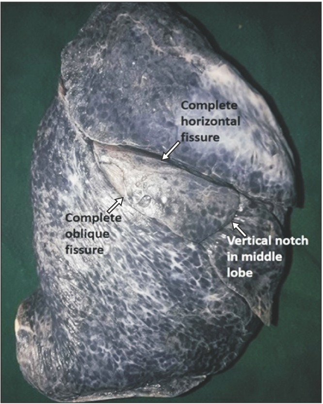
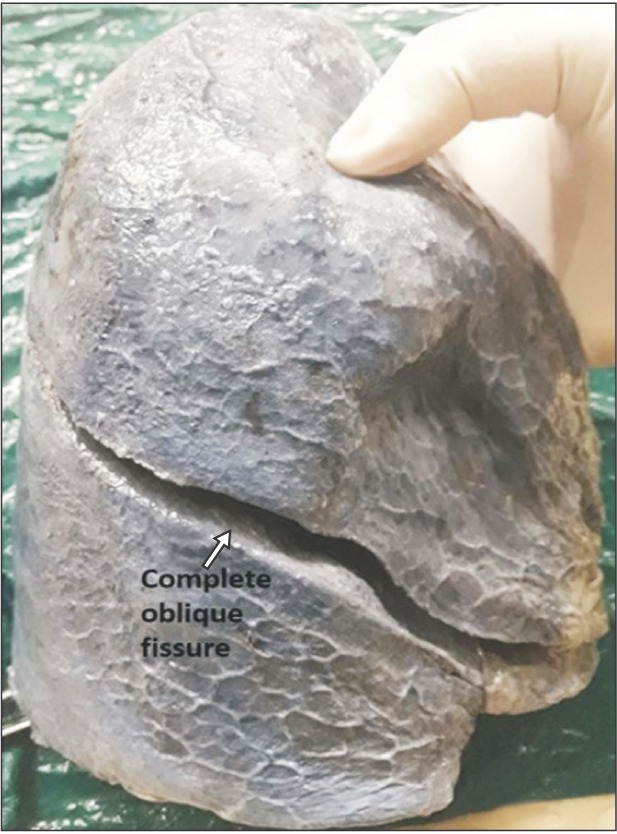
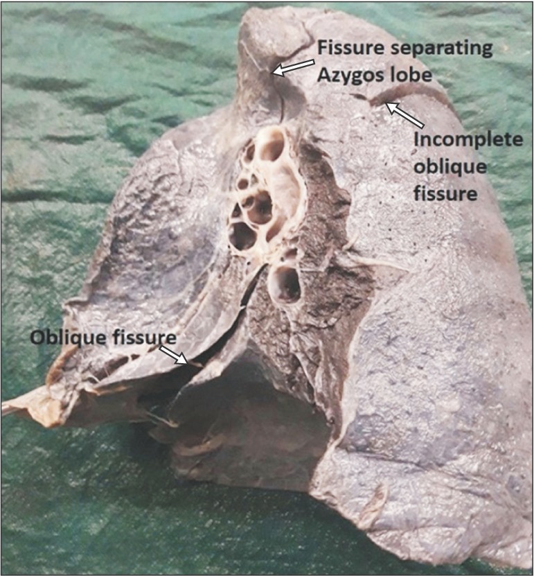
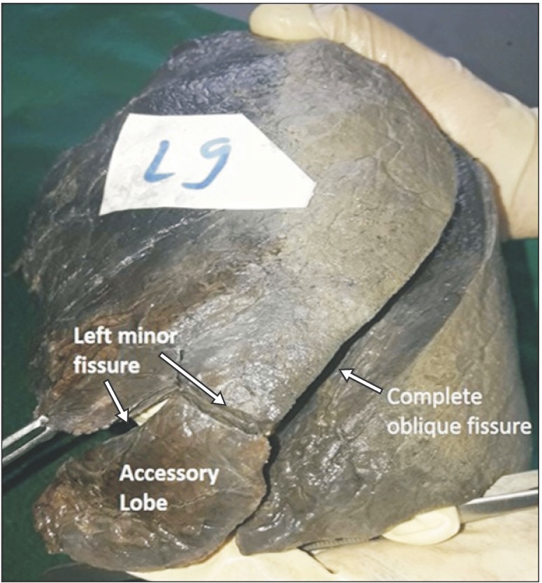
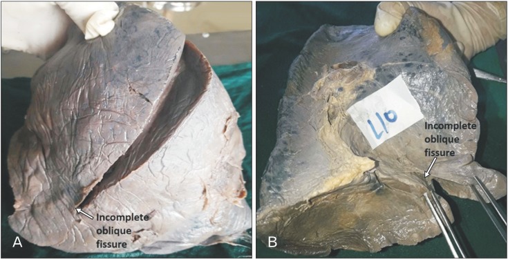
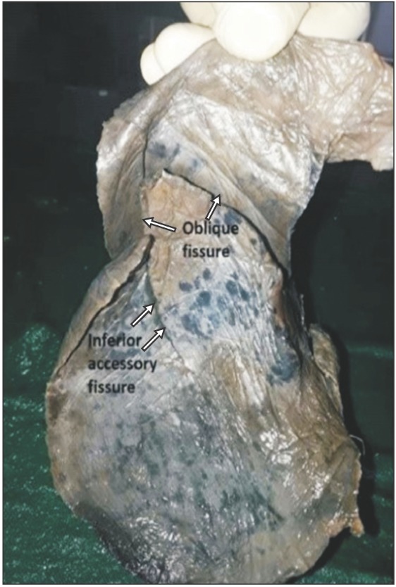
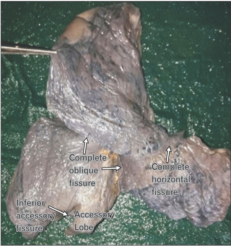
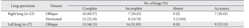
In 23 right lungs; eight were with perfect lobation having complete oblique and horizontal fissure (Fig. 1). Two right lungs had both horizontal and oblique fissures incomplete. Three lungs had complete absence of horizontal fissure (Fig. 2); among which two had complete and one had incomplete oblique fissure, hence only two lobes were present. The complete oblique with incomplete horizontal fissure was found in six specimens while incomplete oblique with complete horizontal fissure was found in four specimens. One right lung with “lobe of the azygos vein” (Fig. 3) was also found. In 27 left lungs; 13 lungs exhibited with complete oblique fissure (Fig. 4) and 14 specimens had incomplete oblique fissure (Fig. 5A, B) with imperfect lobation. None of the right and left lugs showed absence of oblique fissure.
The observations regarding variation in oblique fissures, horizontal fissures, and accessory fissures in both right and left lungs are tabulated in Table 1. The oblique fissure was complete in 16 (69.57%) of right lung and 13 (48.15%) of left lung while it was found to be incomplete in seven out of 23 right lung specimens (30.43%) and 14 out of 27 lung specimens (51.85%). Twelve (52.18%) horizontal fissures of right lung were complete, eight (34.78%) were incomplete and three (13.04%) were absent. The criteria here used to classify the lung fissure incompleteness was the connected lobes by chunk of pulmonary tissue anywhere in the fissure, and those failed to reach the hilum of lung.
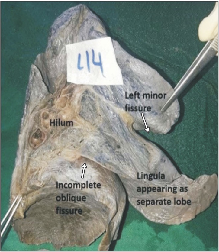
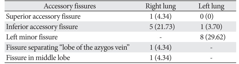
Accessory fissure varied from notch to complete fissure; and exhibited in seven right lungs (Table 1) among them one lung had both SAF (Fig. 6) as well as IAF (Fig. 7). Four right lungs had only IAF, because of which additional lobe (Fig. 8) can be appreciated in inferior lobe. One right lung had accessory fissure in medial surface above the hilum demarcating the “lobe of the azygos vein” (Fig. 3). Interestingly, one right lung had a notch in the costal surface of middle lobe probably representing the demarcation between medial and lateral bronchopulmonary segment (Fig. 1). Nine left lungs (Table 1) were with accessory fissure. Among them IAF was found in one specimen while other eight (Table 2) had LMF (Fig. 9). In two specimens the lingula appeared to be separate lobe (Fig. 9) because of LMF.
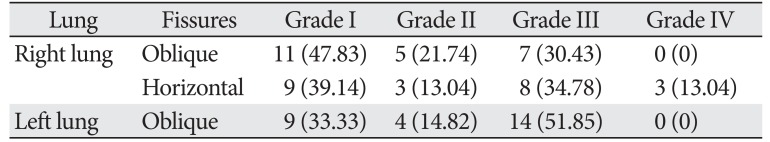
The data in Craig and Walker's classification (Table 3) shows 33.33% of left sided and 47.83% of right sided lung oblique fissure, and 39.14% of horizontal fissure can be classified as grade I. Similarly, 14.82% oblique fissure of left lung, 21.74% oblique fissure of right lung including 13.04% of horizontal fissure of right lung can be categorized as grade II. Grade III fissures were seen in 51.85% and 30.43% of oblique fissures of left and right lungs respectively and 34.78% of horizontal fissures of right lung. Only 13.04% of grade IV horizontal fissure was found. The criteria here used to classify degree of completeness of fissures were the location of pulmonary artery at the base of oblique fissure and extent of varied parenchymal fusion along the floor of fissure. Gradation of fissures is important surgically for easy approach in surgical procedure and to prevent postoperative hemorrhage and complications.
Discussion
When the embryo is approximately 4 weeks old, the respiratory diverticulum (lung bud), an outgrowth from the ventral wall of the foregut, expands caudally into the surrounding mesenchyme and bifurcates into right and left bronchial buds. The right bronchial bud then divides into three secondary bronchi while the left bud into two secondary bronchi. Each lung then develops by a process of repeated dichotomous branching of the secondary bronchi. After several generations of branching, bronchopulmonary segments are formed [11]. In fetal period these bronchopulmonary segments are separated by spaces which later on gets obliterated except along the line of division of principal bronchi to give rise to major (oblique) and minor (horizontal) fissures in fully developed lung. Along these fissures the visceral pleura is reflected and covers individual lobes on all sides [12]. Defective pulmonary development will give rise to variations as encountered in fissures and lobes [13]. Incomplete or absence of oblique and horizontal fissures could be due to a defect in the obliteration of these fissures either completely or incompletely [2]. The monopodial branching of stem bronchi accounts for accessory bronchi and lobes often found in adult lung [14].
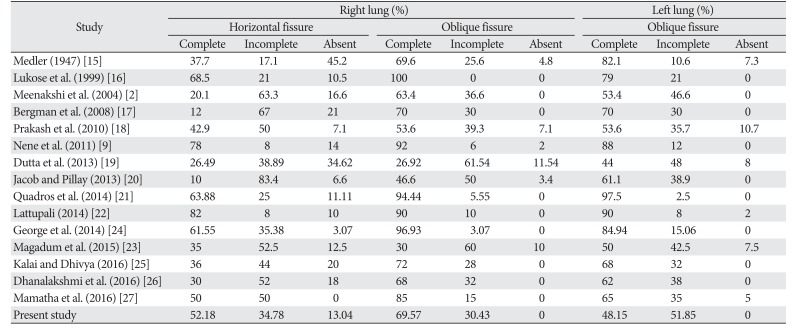
Cadavers are still the best means to study all the domains of anatomy. Various Researchers in different studies of lungs performed on cadavers have reported their findings time and again which is compared with the present study as shown in Tables 4 and 5 [2915161718192021222324252627].
It is observed that absent horizontal fissure is commonly occurring variation as reported by many researchers (Table 4). Only the study of Mamatha et al. [27] did not show its appearance. Incidence of missing horizontal fissure in present study (13.04%) is less than some studies [291517192526] but higher than other studies [16182021222324]. Incomplete horizontal fissure was found highest in Jacob and Pillay's study [20] while least was in Lattupalli's study [22]. In our study it is found to be 34.78%.
Prevalence of absence of oblique fissure in both right and left lung is comparatively less than absence of horizontal fissure in all above studies including present study. Oblique fissure of right lung was reported to be absent in data of other studies [91518192024] while similar to our study remaining researchers [21617212223252627] did not find missing oblique fissures. Incidence of missing oblique fissures of left lung was reported [151819222427] but none of the cases of missing oblique fissures of left lung were reported in remaining studies including present study.
In present study incomplete oblique fissure was more in left lung (51.85%) in comparison to right lung (30.43%). Such findings were similar in most of above mentioned cases except these studies [181920212223]. “Lobe of the azygos vein” beside present study was reported only by Lattupalli [22].
Beside variations in major and minor fissures of lung, clinicians and radiologists must be aware of possibilities of having accessory fissure. Anatomically, an accessory fissure is a cleft of varying depth lined by visceral pleura. Radiographically it appears as a thin white line, resembling the major and minor fissure, except for location. The line can be mistaken for an interlobar fissure, scar and wall of a bulla or for pleural line made visible by pneumothorax [4].
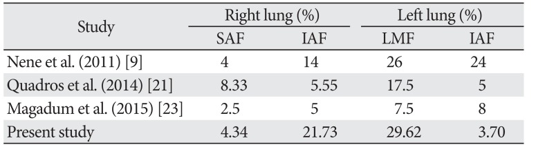
As shown in Table 5, in present study LMF (29.62%) was found to be most frequently occurring accessory fissure which is similar to study of Nene et al. (26%) [9], Quadros et al. (17.5%) [21], and George et al. (7.5%) [24]. According to Nene et al. [9], Quadros et al. [21], and George et al. [24], SAF of right lungs were 4%, 33%, and 2.5% respectively and in present study it was 4.34%. Study of Quadros et al. [21] showed the higher incidence of IAF in right lung (5.55%) than in left lung (5%) which is similar to our study i.e. 21.73% in right lung and 3.70% in left lung, while Nene et al. [9] had contrasting finding, more percentage of IAF in left lung (24%) than in right lung (14%). George et al. [24] did not find IAF in left lung and incidence in right lung of his study was 5%. Studies of Prakash et al. [18], Meenakshi et al. [2], and Kalai and Dhivya [25] did not report any accessory fissures.
According to Godwin and Tarver [4], besides SAF, IAF, and LMF other accessory fissures are also occurring in other locations. The more common locations are between medial and lateral segments of the middle lobe and between anterior and lateral segments of the lower lobes. Sometimes fissures occur within a bronchopulmonary segment, usually separating subsegments, most commonly in the middle lobe or the posterior segment of the left lower lobe [4]. In present study a vertical notch was observed in middle lobe of one right lung, probably separating the medial and lateral bronchopulmonary segments of the lobe. Such finding was not shown in any literatures available to our best of knowledge.
The nature of fissure is of great importance in planning pulmonary surgeries. In order to provide a frame work for description of operative technique and to allow meaningful comparison between different surgical series, Craig and Walker [10] have proposed a fissural classification based on both the degree of completeness of the fissures and the location of the pulmonary artery at the base of the oblique fissure [2]. Gradation of fissures is important surgically for easy approach in surgical procedure and to prevent postoperative haemorrhage and complications.
In cases of incomplete fissures where parenchymal fusion is present more dissections has to be performed to reach the bronchi and pulmonary arteries during surgical resections leading to more haemorrhage and postoperative complications. An incomplete fissure may alter the spread of disease within the lung and is also a cause for postoperative air leakage [10]. Similarly, accessory fissures in patients with endobronchial lesion, might alter the usual pattern of lung collapse and pose difficulty in diagnosing a lesion and its extent. Often these accessory fissures act as a barrier to spread of infection, creating a sharply marginated pneumonia, which can wrongly be interpreted as atelectasis or consolidation [4]. An anomalous fissure can be mistaken for a lung lesion or an atypical appearance of pleural effusion.
As the fissures form the boundaries for the lobes of the lungs, knowledge of their position is necessary for the appreciation of lobar anatomy and thus for locating the bronchopulmonary segments which is significant both anatomically and clinically. The lobes of lungs show partial fusion as a result of incomplete pulmonary fissures. The results and their comparison with the previous works show that there is a wide range of difference in occurrence of oblique, horizontal and accessory fissures. In present study, common finding is complete oblique and horizontal fissures in right lung and incomplete oblique fissure in left lung. Accessory fissure varied from notch to prominent fissure often separating an accessory lobe. LMF of left lung, which separates the lingula from upper lobe is frequently appearing accessory fissure. SAF and IAF is more common in right lung in comparison to left lung. The wide range of variation in occurrence of oblique, horizontal and accessory fissures might be due to genetic and environmental factors during its development.

 XML Download