Dear Editor:
Hypertrichosis is the excessive growth of mature hair on non-androgen-dependent areas of the body that is abnormal for age, sex, or race1. It can be generalized or localized, with variable etiologies ranging from genodermatoses to underlying tumorous conditions. Localized acquired hypertrichosis was usually reported in cases of repeated trauma, friction, irritation, or inflammation2. We herein report a rare case of plexiform schwannoma with localized acquired hypertrichosis.
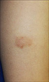
A 5-year-old boy presented with a solitary brown nodule on his left leg that had been present since birth. The lesion began to grow several months before, and localized hypertrichosis overlying the nodule was also observed after the lesion had grown (Fig. 1). No other skin lesions were observed, and the patient was otherwise healthy with no developmental problems. There was no family history of localized hypertrichosis. Skin biopsy was performed from the solitary nodule, and histopathological finding showed multiple variable-sized, encapsulated, and interconnected nodules composed of spindle cells with focal palisading nuclei (Fig. 2A, B). In addition, the tumor cells in the nodules were strongly stained with S-100 protein (Fig. 2C). Only the capsules of the nodules were stained with epithelial membrane antigen (Fig. 2D). Neurofilament staining of the cells in nodules was negative (Fig. 2E). Based on the histopathological findings, plexiform schwannoma was diagnosed. The lesion was surgically excised, and no recurrence was observed.
Plexiform schwannoma is a rare benign tumor of the nerve sheath. It usually occurs in young adults and presents mostly as a slowly growing asymptomatic solitary nodule on the trunk, head and neck, and upper extremities. It can be accompanied with size enlargement or pain3. However, to the best of our knowledge, no case associated with localized hypertrichosis has been reported. Neurofibroma, another benign tumor of the nerve sheath, has been reported with localized hypertrichosis14. Although the exact pathomechanism of localized hypertrichosis with neurofibroma has not been clarified yet, some hypotheses have been suggested for hypertrichosis. Growth factors, such as epidermal growth factor, vascular endothelial growth factor, platelet-derived growth factor, and bone morphogenic protein, have been determined to play a role in creating an environment that affects the anagen phase of the hair follicle cycle5. Thereby, the prolongation of the anagen phase, due to the interaction between hair follicles and various growth factors derived from the tumor cells, has been suggested as the main mechanism in the pathogenesis of localized hypertrichosis overlying tumors14.
In this case, the patient had no history of trauma, inflammation, drug, or vaccination. He only had a slowgrowing plexiform schwannoma at the site of the hypertrichosis, which occurred after the tumor had grown. Thus, we think that the changes in the hair follicle cycle due to the various growth factors derived from the tumor could be suggested as the possible pathogenesis.
Figures and Tables
Fig. 2
(A) Multiple variable-sized, encapsulated, and interconnected nodules (H&E, ×40). (B) Nodules composed of spindle cells with focally palisading nuclei (H&E, ×100). (C) Tumor cells in the nodules strongly stained for S-100 protein (S-100 protein staining, ×100). (D) The capsules of the nodules stained for epithelial membrane antigen (EMA, ×200). (E) Neurofilament staining of the cells in the nodules was negative (neurofilament staining, ×100).

References
1. Cho E, Kim HS, Lee JY, Kim HO, Park YM. Localized hypertrichosis overlying neurofibroma. Int J Dermatol. 2013; 52:1623–1624.

2. Camacho-Martinez FM. Dermatology. 3rd ed. Philadelphia: Elsevier Saunders;2012. p. 1115–1127.
3. Ko JY, Kim JE, Kim YH, Ro YS. Cutaneous plexiform schwannomas in a patient with neurofibromatosis type 2. Ann Dermatol. 2009; 21:402–405.


 PDF
PDF ePub
ePub Citation
 Print
Print


 XML Download