

 PDF
PDF ePub
ePub Citation
 Print
Print


Dear Editor:
Granuloma annulare (GA) is a common cutaneous granulomatous disease of unknown etiology, associated with several systemic conditions including diabetes mellitus, thyroid disease, dyslipidemia, and malignancy1. Although an association with hepatocellular carcinoma (HCC) had been suggested in one case, a true paraneoplastic course could not be established because of the patient's demise a couple of months after presentation2. We report a rare case of paraneoplastic generalized GA in a patient with HCC.
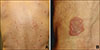
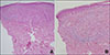
A 71-year-old man presented with a 3-year history of erythematous papules and plaques on his back and upper extremities (Fig. 1) without any itching or pain. He had no significant personal or family history of skin disease. He was diagnosed 3 years 6 months ago, as having intermediate-stage HCC with underlying liver disease including alcoholic liver cirrhosis and resolved hepatitis B virus infection. He was administered several anti-tumor treatments, viz., transarterial chemoembolization, stereotactic radiation therapy, and a tyrosine protein kinase inhibitor (sorafenib). Sorafenib treatment was initiated 1 year 6 months ago, due to progression of HCC into the advanced stage. It resulted in temporary complete remission a year ago (Supplementary Fig. 1). However, he soon showed progressive disease. The patient currently refuses to receive any further active treatment and seeks only supportive care. Interestingly, he noted that his cutaneous lesions diminished during the phase of complete remission of HCC. A skin biopsy revealed palisading granulomatous inflammation with histiocytic infiltration surrounding collagen bundles in the dermis (Fig. 2). Based on these findings, the patient was diagnosed as having generalized GA and was subsequently treated with 0.25% topical prednicarbate ointment.
GA is a benign granulomatous skin disease that usually presents as annular skin-colored to erythematous papules localized to the dorsal aspects of the hands or feet. There are four clinical variants: localized, generalized, subcutaneous and perforating. According to a multicenter study analyzed 54 patients with generalized GA, generalized GA usually occurred on the trunk and extremities. These can be differentiated from the localized type in that they were observed in an older age group, had widespread distribution, and were refractory to therapy3. Several investigators have suggested a relationship between GA and other systemic diseases, most commonly diabetes mellitus. Additionally, GA is reported to be associated with malignancies, including hematopoietic cancers and adenocarcinomas; however, an association between GA and liver disease has hardly been reported14. Mestre et al.2 reported a case of generalized GA in a patient with HCC, although the authors failed to establish a temporal relationship between GA and the malignancy owing to the patient's demise. The case of Mestre et al.2 showed mixed papules and annular plaques, similar to the present case. The paraneoplastic variant of GA shows a poor response to conventional GA treatment, although it resolves spontaneously after removal of the underlying neoplasm5. The possible mechanism of paraneoplastic GA includes tumor antigens triggering an immunological response, which results in a granulomatous reaction25.
In conclusion, we report the second case of generalized GA in a patient with HCC, in whom GA demonstrated a paraneoplastic course, as lesions diminished during the temporary phase of remission of the HCC treated with sorafenib. Therefore, the cutaneous lesions were presumably caused by a paraneoplastic syndrome from an internal malignancy. In elderly patients demonstrating generalized GA, dermatologists must consider underlying malignancy including HCC.
 XML Download