

 PDF
PDF ePub
ePub Citation
 Print
Print


INTRODUCTION
Chronic lymphocytic leukemia (CLL) is a malignant lymphoproliferative disorder characterized by progressive accumulation of leukemic cells in the blood, bone marrow, and lymphoid tissues1. Any other organ can be involved, and the skin is a common extra-nodal site for the accumulation of leukemic cells1. Skin eruptions are also common in patients with CLL, and these lesions can develop as non-specific cutaneous reactions without cutaneous infiltration of leukemic cells2.
An “exaggerated reaction to an insect bite” has been reported as a non-specific cutaneous reaction that is rarely observed in patients with CLL3. The clinical features include recurrent pruritic erythematous patches, papules, plaques, and bullae, which are similar to the clinical features of insect bites or autoimmune bullous diseases2. However, the reactions have been described as “insect bite-like reactions” or “eosinophilic eruption of hematoproliferative diseases” because the lesions are often triggered without a preceding arthropod bite2. We report a case of an insect bite-like reaction with bullous lesions in a 74-year-old man with CLL. The study was approved by the Institutional Review Board of the Gangnam Severance Hospital (IRB no. 3-2016-0273). Informed consent was obtained from the patient prior to this report. We received the patient's consent form about publishing all photographic materials.
CASE REPORT
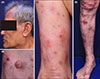
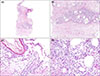
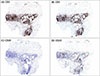
A 74-year-old an presented with a 2-year history of pruritic recurrent erythematous patches and bullae on the whole body. The patient had been diagnosed with CLL in 2015. Immunophenotyping with the bone marrow biopsy showed positive for CD5, CD20, CD23 in leukemic cells. He had achieved complete remission after 2 months of chemotherapy with chlorambucil, fludarabine, cyclophosphamide, and rituximab. The skin lesions developed 1 month after the complete remission of CLL. The skin lesions had responded to oral methylprednisolone, but tended to relapse with tapering of methylprdnisolone, and the patient experienced worsening of the lesions and itching sensation 6 months before his presentation. A physical examination revealed erythematous patches and bullae on the whole body (Fig. 1), and we suspected a diagnosis of bullous pemphigoid. Laboratory test revealed an elevated eosinophil count (26.6%, 2,020/µl). Skin biopsy at the right knee revealed mild spongiosis of the epidermis, subepidermal bulla, and edema at the papillary dermis. Lymphocytic infiltration with eosinophils was found throughout the dermis and subcutaneous fat layer (Fig. 2). Immunohistochemical staining of the infiltrated cells showed positive reactions for CD3, CD5 and negative for CD20, CD23 (Fig. 3). Direct and indirect immunofluorescence revealed negative results. The diagnosis of CLL-associated insect bite-like reactions was made based on the histopathological features and immunofluorescence results.
The skin lesions resolved 3 months after treatment with oral methylprednisolone (8~12 mg), dapsone (50 mg) and topical diflucortolone valerate. The patient has remained in remission with low-dose methylprednisolone (4 mg) and dapsone (25 mg) during a 6-month follow-up.
DISCUSSION
In 1965, Weed3 first reported an exaggerated reaction to an insect bite in a patient with CLL, who developed induration, edema, erythema, and bullae with intense pruritus at the site of the insect bite. He suggested that this reaction was a delayed hypersensitivity reaction to the insect bites, based on the patient's altered immune response caused by the CLL3. However, more recent studies have frequently reported insect bite-like reactions in patients with CLL without the patient's recalling an insect bites2. Furthermore, the lesions were often not restricted to exposed sites and did not exhibit seasonal variations, which indicate that the lesions might not be triggered by an insect bite2. Barzilai et al.4 have suggested the term “insect bite-like reaction” for this phenomenon, which was further defined as “eosinophilic dermatosis of myeloproliferative disease” by Byrd et al.5 These authors also proposed diagnostic criteria: (1) pruritic papules, nodules, and/or vesiculobullous eruptions that were resistant to conservative management; (2) histopathologically confirmed eosinophil-rich dermal lymphohistiocytic infiltration at the superficial and deep dermis; (3) exclusion of other causes of tissue eosinophilia; and (4) a pre-existing diagnosis of a hematological malignancy5. Insect bite-like reaction with bullous eruption shows similar clinical and histologic features of bullos pemphigoid. Thus, direct and indirect immunofluorescence examinations are mandatory to make correct diagnosis.
The pathogenesis of insect bite-like reactions remains unclear. The skin lesions have been considered as a non-specific cutaneous reaction to certain stimuli in patients with hematologic diseses, not a specific reaction by leukemic cells. A recent study using in situ hybridization analysis demonstrated that neoplastic CLL cells were observed within insect bite-like skin lesions, suggesting they may be specific skin lesions, rather than non-specific cutaneous skin reactions67. In our case, immunohistochemical stain showed negative for CD20 and CD 23 in skin, which are highly expressed in neoplastic CLL cells and also detected in our patient's bone marrow biopsy, indicating that the lesions are not related to skin infiltrates of the neoplastic CLL cells8. In fact, insect bites, drugs, chemoimmunotherapy, and pyogenic infections can trigger non-specific eosinophilic eruptions in patients with CLL2. The altered immune response in patients with hematological disease can increase the secretion of interleukin 4 (IL-4) and IL-5, which stimulates eosinophilic skin infiltration4.
The development of skin lesions are usually not related with the course or activity of CLL as our case2. Bairey et al.2 have reported that the skin eruptions can occur before diagnosis of CLL or after the end of chemotherapy. Oral glucocorticoids, intravenous immunoglobulin, and dapsone have been reported to be effective for controlling the lesions49. Re-initiation of chemotherapy may also help improve the skin lesions in some of patients10. Our patient showed remarkable improvement after receiving combination therapy with oral methylprednisolone and dapsone. In conclusion, insect bite-like reaction with bullous lesions should be considered, particulary when a patient with CLL shows chronic insect bite-like or bullous pemphigoid-like lesions.
 XML Download