

 PDF
PDF ePub
ePub Citation
 Print
Print


INTRODUCTION
Mucinous nevus is a rare disorder classified as either a cutaneous mucinosis or a connective tissue nevus12. The condition was first described by Redondo Bellón et al.1 in 1993. Clinically, asymptomatic grouped papules or plaques grow to form a verrucous or nevoid feature exhibiting a unilateral or zosteriform distribution34. The nevus usually develops on the trunk at birth or in early adulthood34. Histologically, the nevus is characterized by mucin deposits localized to the superficial dermis34.
CASE REPORT
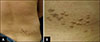
A previously healthy 34-year-old Korean male presented with asymptomatic grouped gray-brown papules and confluent plaques exhibiting a zosteriform distribution on his right lower back (Fig. 1). The skin lesions had commenced in childhood, gradually coalesced, and grew slowly. In our patient, pigmentary abnormalities as like freckles except for skin lesions of the right lower back were not observed. He had neither any past medical problem nor a family history of similar lesions and pigmentary abnormalities. He denied any trauma.
Histological examination revealed an acanthosis with elongated rete ridges and amorphous materials associated with loosely separated collagen fibers in the papillary dermis (Fig. 2A). The amorphous materials stained with alcian blue at pH 2.5 (Fig. 2B); this confirmed a mucin deposit limited to the papillary dermis. Verhoeff-van Gieson staining revealed that the numbers of elastic fibers in the papillary dermis were reduced in the regions of mucin deposition (Fig. 2C).
This clinicopathological analysis enabled us to diagnose a mucinous nevus. Our patient decided to allow us to observe the lesion; no treatment was performed.
DISCUSSION
Mucinous nevus is an uncommon entity initially described by Redondo Bellón et al.1 in 1993 and classified as either a cutaneous mucinosis or a connective tissue nevus12. The cutaneous mucinoses are a heterogeneous group of diseases in which abnormal amounts of mucin are deposited in the skin6. Connective tissue nevi are hamartomas with unusual levels (excesses or deficiencies) of collagen, elastin, and/or proteoglycans2.
The term “mucinous nevus” refers to the its nevoid appearance and the characteristic pattern of mucin deposits in the papillary dermis2613. Clinically, mucinous nevi present as asymptomatic, multiple skin-colored to brownish papules or plaques; separate lesions coalesce and then grow to form a verrucous or nevoid feature with a unilateral or zosteriform pattern346. It usually develops at birth or in early adulthood613. The principal site is the trunk, including the back36. The male:female ratio is 5:1; the reason is not clear5. To date, there are two reports of familial mucinous nevus613. However, there was no report about the genetic abnormality as like mosaicism. Histologically, mucinous nevus is characterized by diffuse band-like mucin deposits in the uppermost portion of the dermis1234. The mucin is thought to be composed of hyaluronic acid staining positively with alcian blue at pH 2.5 but not staining at pH 0.51415. The origin of the mucin remains unknown14, but is presumed to be attributable to a primary metabolic process (such as overproduction) rather than a secondary catabolic process2. Mucinous nevi are divided into two histopathological types depending on whether epidermal changes are present; these are connective tissue nevi of the proteoglycan (CTNP) type and the combined epidermal-CTNP type5. The epidermis is normal, in the CTNP type but, in the combined epidermal-CTNP type, exhibits hyperkeratosis and acanthosis with elongation of the rete ridges2. Our case featured an epidermal change; thus, we diagnosed the combined epidermal-CTNP type of mucinous nevus.
Both an epidermal nevus and nevus lipomatosus superficialis exhibit nevoid features similar to those of a mucinous nevus. It is difficult, therefore, to clinically distinguish among the conditions. Histological data are necessary. Histologically, an epidermal nevus and nevus lipomatosus superficialis can be distinguished from a mucinous nevus; only the latter exhibits mucin deposits in the papillary dermis.
However, such mucin deposition is also observed in cutaneous mucinosis of infancy, but is very superficial and appears to be hugged by the epidermis5. Clinically, cutaneous mucinosis of infancy presents as scattered small papules unlike mucinous nevus.
Mucinous nevi do not require treatment (except for cosmetic purposes); the nevi are benign3. Surgical excision, scalpel dermabrasion, and carbon dioxide laser treatment are possible5. Surgical excision is not indicated if several discrete lesions are evident5. Mucinous nevus of the CTNP type was treated via scalpel dermabrasion, but scarring developed 1 year later16. There is one report of mucinous nevus of the combined epidermal-CTNP type which did not recur after carbon dioxide laser vaporization5. Our patient did not voice any cosmetic concern; thus, we decided to simply observe the lesions.
To the best of our knowledge, only 25 cases of mucinous nevi have been reported in the English-language literature3567891011 and only one in the Korean literature12. The principal location is the trunk including the back. Approximately 80% of all cases were reported in males and about 30% of all cases present at birth.
Herein, we report a rare case of mucinous nevus of the combined epidermal-CTNP type.

 XML Download