

 PDF
PDF ePub
ePub Citation
 Print
Print


Introduction
Coronary artery is one among the most important arteries and even a short term blockage of it might result in life threatening myocardial infarction. Many of the variations of coronary artery may not be hazardous and may go unnoticed throughout life. However, some variations may affect the functioning of the heart seriously. They may also cause problems during stent placements and some diagnostic and therapeutic procedures. Right coronary artery (RCA) may show variations in its origin, course, termination, and distribution. Some of its reported variations include its absence, being larger than usual, origin from pulmonary trunk, and giving additional branches. A sound knowledge of all possible variations of coronary arteries is useful for cardiologists and radiologists for successful diagnosis and treatment of the coronary artery disease. Here, an extremely rare variation of RCA is being reported.
Case Report
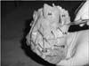
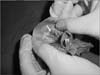
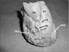
During routine dissection classes for medical undergraduates, an early trifurcation of the RCA was observed in an adult male cadaver aged 65 years approximately. The RCA had a normal origin from the anterior aortic sinus with a single opening. After a course of 2 mm, it trifurcated. Among the three branches, the first was the right conus artery, the second branch was a huge right ventricular branch and the third was the main continuation of the RCA. The right conus artery entered the myocardium after a course of 1.5 cm. The huge ventricular branch had the same diameter as the main continuation of the RCA and it ran downwards and to the left on the middle of the anterior surface of the right ventricle, bridged by some myocardial fibers here and there. It ran down almost up to the apex of the heart, and then entered the myocardium. The large ventricular branch supplied the myocardium of the right ventricle. The right marginal artery was absent. The anterior interventricular artery had a normal size and distribution, but it was covered by fat and some myocardial fibres in the anterior interventricular sulcus. There were no other vascular variations of the heart apart from this. There was no notable change in the thickness of the walls (ventricles) of the heart. The trifurcation and the three branches of RCA have been shown in Figs. 1, 2, 3.
Discussion
Variations of origin and branching pattern of RCA have been well documented. It usually arises from the anterior aortic sinus of Valsalva. However, in some cases, it might take its origin from upper part of ascending aorta as ectopic origin [1]. Its origin from the pulmonary trunk has also been reported [2] and such an anomalous origin does not usually cause any functional problems. Origin of RCA along with left coronary artery (LCA) as a common trunk has been documented in the literature [3]. In rare cases, RCA originates as a septal branch of LCA [4]. Some cases of origin of RCA from left anterior descending artery have also been documented [5]. In some cases, the RCA duplicates or splits. This variant of RCA can predispose the heart for myocardial infarctions [67]. In some individuals, the RCA is super dominant and can replace the circumflex artery [8]. A thorough literature survey with regards to the current variation did not reveal any report on trifurcation of the RCA though the trifurcation and associated complications of LCA have been well documented [9]. Possibly, this could be the first report on such a trifurcation of RCA very close to its origin. Radiologic diagnosis, percutaneous approach and stent placing is a challenge in cases of trifurcation of coronary arteries [10]. The current case is unique due to the trifurcation of the RCA and also for the presence of the huge ventricular branch passing down parallel to the left anterior descending artery towards the apex of the heart. The site of trifurcation of RCA may be prone for coronary artery disease and can be really challenging in placing the stent. Trifurcation of RCA might not cause any functional disturbances to the heart as it supplied all the areas which are usually supplied by the RCA. To the best of my knowledge, this could be the first report on the trifurcation of RCA.
 XML Download