

 PDF
PDF ePub
ePub Citation
 Print
Print


Introduction
Osteochondromas are the commonest benign tumors of the skeleton; develop in the periosteum as small cartilaginous nodules [1]. They are developmental in origin rather than the true neoplasm. They are estimated to constitute around 20%–50% of all benign bone tumors and are found to exist in the form of solitary or multiple, pedunculated or sessile. Solitary osteochondromas develop as a bony projection with cartilaginous cap. They are typically observed in the long bones as slow growing projections from the metaphysis of the long bones, and stop growing once the fusion of growth plate is complete. They are also found to occur in the bones of axial skeleton, flat bones of skull and facial bones [1]. In the present case we report a case of solitary osteochondroma in the body of the right pubic bone.
Case Report
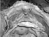
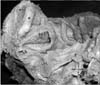
During a regular dissection class for medical undergraduates, we came across a bony nodule in the body of the right pubic bone. It was observed in an approximately 75-year-old formalin embalmed female cadaver of South Indian origin. It was found to be a sessile osteochondroma and was situated in the posterior surface of the body of the right pubic bone, very close to the pubic symphysis (Figs. 1, 2). It projected backwards, into the pelvic cavity. Histopathology of this sessile nodule confirmed the typical features of the osteochondroma. The bony nodule presented marrow (trabecular bone) and cortical bone which were found to be continuous with the parent bone. Further this bony nodule was covered by a cartilaginous cap (Fig. 3). Since the body was donated to the institution for teaching and research purpose, there are no ethical issues in reporting the present finding.
Discussion
The observed finding of osteochondroma on the body of pubic bone is incidental and is of academic interest. It was noted during dissection classes for undergraduate students. Incidence of pubic bone osteochondroma is very rare and is seldom reported in the literature. Hence, we report the present case with review of literature and discuss its clinical implications.
In general, the solitary osteochondromas are found in the bones that are performed in the endochondral ossification. Males are predominantly affected than females with a ratio of 1.74–1 [2]. They are frequently observed in the long tubular bones (60%) and short tubular bones (10%) [3]. Rarely found to occur in in the axial skeleton, flat bones of skull and facial bones [1]. Osteochondroma in flat bones usually develops in the parts which are most recently formed from cartilage, for example in the scapula along the inner border; in ilium along the iliac crest and around the acetabulum [2]. Osteochondromas have been documented in all the pelvic bones and its incidence is found to be 5% among all osteochondromas [3]. Kumar et al. [4] have reported a case of osteochondroma on ileum bone. There are cases of osteochondroma of pubic symphysis causing sexual disturbances [5]. Buzon [6] has reported two cases of osteochondroma in the os pubis. In both the cases they have found the bony lump projecting from the anterior surface of the right pubis. In a 29-year-old man, osteochondroma was found on the superior ramus of the left pubic bone [7]. Recently, in an 18-year-old young female, it was found on the ramus of right pubis [8]. No evidence of recurrence was noted in above two cases after excision. Contrary to previous reports, we observed osteochondroma on the posterior surface of the body of the pubis, projecting into the pelvic cavity. To the best of our knowledge, this is the first report in literature.
Osteochondromas are usually asymptomatic and present as characteristic pain less masses. However, they become symptomatic when they compress the vascular and neurological structures [9]. In addition, the pelvic osteochondromas may compress the urogenital structures. Rarely, the excess bony growths may become chondrosarcomas [10]. The current case of the osteochondroma on pubic bone body might put pressure on the urinary bladder and urethra resulting in increased frequency in micturition and might result in sexual problem. When it happens in relatively younger individual, it might cause complications during parturition by reducing the dimensions of the pelvic cavity. The knowledge of rare possibility of this type of bone growth on the dorsal aspect of pubis could be important to orthopedic surgeons during the treatment of fracture/dislocations of the pubis. It could be useful also to andrologists and urologists as it could lead to sexual and urinary problems. Though occurrence of osteochondromas is common in the pelvic bones, its location on the posterior surface of the body of the pubis, projecting towards the pelvic cavity is remarkable. The knowledge the incidence of such unusual osteochondromas may be clinically important during the diagnosis and planning of treatment when they are symptomatic.

 XML Download