

 PDF
PDF ePub
ePub Citation
 Print
Print


Ho Min Lee, M.D., Jong Pil Kim, M.D. , Suk Kang, M.D., Young Sung Kim, M.D., Min Young Lee, M.D.
, Suk Kang, M.D., Young Sung Kim, M.D., Min Young Lee, M.D.
Abstract
Purpose
Pseudoepiphysis originates from the secondary ossification center of the non-ossification end during the normal pediatric growth process. It is not uncommonly found in the course of metacarpal and metatarsal ossification. We investigated the radiologic prevalence and features of pseudoepiphysis in normal Korean children.
Materials and Methods
Sex and age distribution following radiologic prevalence as well as the features of metacarpal pseudoepiphysis of 2,320 Korean children aged below 15 years of age and younger who underwent hand radiography between January 2009 and February 2016 were analyzed.
Results
A total of 304 out of 2,320 patients had pseudoepiphysis on metacarpal bone, which is a prevalence of 13.1%. Male showed higher prevalence (16.6% for male and 10.5% for female). The peak age was 11 years for boys and 5 years for girls. The first metacarpal bone was most prevalent, with 9.6% of the total population, followed by the second metacarpal bone (5.2%) and fifth metacarpal bone (2.5%). The prevalence of single pseudoepiphysis was 9.4%, and that of multiple pseudoepiphysis was 3.7%. The prevalence of incomplete pseudoepiphysis was 8.9% and was higher than complete pseudoepiphysis (5.6%).
Figures and Tables
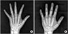 | Figure 1(A) Anteroposterior plain radiography of an 8-year-old female showing complete pseudoepiphysis (arrow) at the base of the second metacarpal bone. Radiolucent pseudoepiphyseal line crosses both the radial and ulnar sides of metacarpal cortex transversely. (B) Incomplete pseudoepiphysis (arrow) presenting at the second metacarpal base on a 6-year-old male radiography, as partial cortical discontinuity.
|
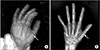 | Figure 2(A) Plain radiography of a 4-year-old male shows single pseudoepiphysis (arrow), as complete pseudoepiphysis of first metacarpal bone. (B) Radiography of an 11-year-old female showing multiple pseudoepiphysis (arrows) at three sites, each on the first, second, and fifth metacarpal bones. In the case, every pseudoepiphysis manifested as an incomplete pseudoepiphysis.
|
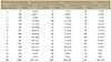
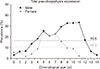 | Figure 3Male patients show higher prevalence of pseudoepiphysis, except for those aged younger than 1 years. An 11-year-old male and a 5-year-old female show the highest prevalence in each sex group. Dotted line shows a value of total pseudoepiphyseal prevalence of each sex.
|
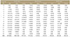
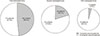 | Figure 4Circular graph shows the proportion of complete and incomplete pseudoepiphysis by the site of expression. Total prevalence of each complete pseudoepiphysis was 5.0%, 4.0%, and 2.4% in the first, second, and fifth metacarpal bone, respectively, and each incomplete pseudoepiphyseal prevalence was 4.7%, 1.2%, and 0.1%.
|
 | Figure 5Anteroposterior plain radiography of a 10-year-old female shows transverse radio-opaque line (arrows) on the distal end of the first metacarpal bone.
|
References
1. Scheuer L, Black S. Developmental juvenile osteology. London: Academic Press;2000. p. 308–340.
2. Nakashima T, Furukawa H. A rare case of complete proximal epiphyses (so-called pseudoepiphyses) of the metacarpal and metatarsal bones in the human. Ann Anat. 1997; 179:549–551.

3. Anderson T. Additional manal and pedal epiphyseal centres. Ann Anat. 2001; 183:485–488.
4. Lee MMC, Garn SM. Pseudoepiphysis or notches in the non-epiphyseal end of metacarpal bones in healthy children. Anat Rec. 1967; 159:263–272.
5. Ogden JA, Ganey TM, Light TR, Greene TL, Belsole RJ. Nonepiphyseal ossification and pseudoepiphysis formation. J Pediatr Orthop. 1994; 14:78–82.
6. Levine E. Notches in the non-epiphyseal ends of the metacarpals and phalanges in children of four South African populations. Am J Phys Anthropol. 1972; 36:407–415.
7. Limb D, Loughenbury PR. The prevalence of pseudoepiphyses in the metacarpals of the growing hand. J Hand Surg Eur Vol. 2012; 37:678–681.
8. Dreizen S, Spirakis CN, Stone RE. The distribution and disposition of anomalous notches in the non-epiphyseal ends of human metacarpal shafts. Am J Phys Anthropol. 1965; 23:181–187.
9. Nesbitt R. Human osteogeny. London: T Wood;1736.
10. Haines RW. The pseudoepiphysis of the first metacarpal of man. J Anat. 1974; 117:145–158.
11. Keats TE, Harrison RB. The epiphyseal spur. Skeletal Radiol. 1980; 5:175–177.
12. Lachman E. Pseudo-epiphyses in hand and foot. Am J Roentgenol Radium Ther Nucl Med. 1953; 70:149–151.
13. Park S, Yang WY, Ryu KN. Study of hand anomaly and bone age in Down syndrome with Korean. J Korean Soc Plast Reconstr Surg. 1999; 26:460–465.
14. Frazer JES. The anatomy of the human skeleton. London: J. & A. Churchill;1940. p. 118.
15. Greulich WW, Pyle SI. Radiographic atlas of skeletal development of hand and wrist. Am J Hum Genet. 1959; 11:282–283.
16. Hefke HW. Roentgenologic study of anomalies of the hands in one hundred cases of mongolism. Am J Dis Child. 1940; 60:1319–1323.
 XML Download