Abstract
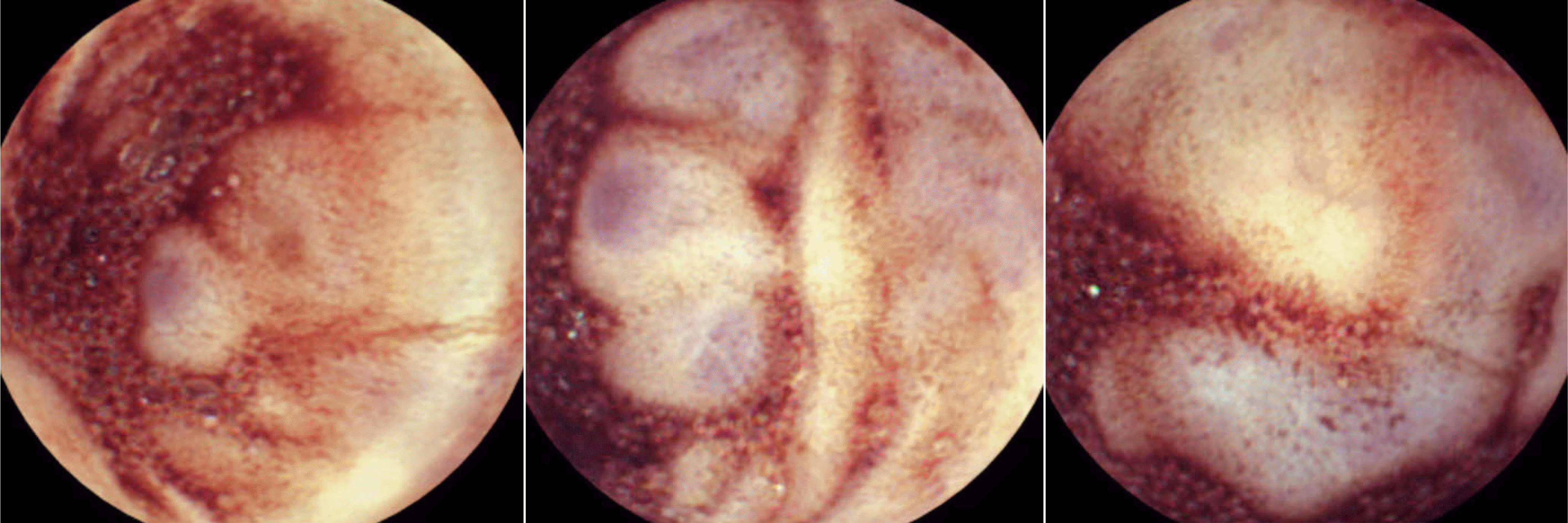
We report a case of bleeding ileal varices associated with intraabdominal adhesions after colectomy which was successfully diagnosed using capsule endoscopy. A 77-year-old woman visited the emergency department for several episodes of melena. She had a medical history of neoadjuvant chemoradiation therapy and subsequent surgery for rectal cancer 6 years previously. Conventional diagnostic examinations including upper endoscopy, colonoscopy, and abdominal computed tomography could not detect any bleeding focus, however, following capsule endoscopy revealed venous dilatations with some fresh blood in the distal ileum, indicating bleeding ileal varices. The patient underwent exploratory laparotomy and the affected ileum was successfully resected. No further gastrointestinal bleeding occurred during the 6 months follow-up. Small intestinal varices are important differential for obscure gastrointestinal bleeding especially in patients with a history of abdominal surgery in the absence of liver cirrhosis, and capsule endoscopy can be a good option for diagnosing small intestinal varices.
Go to : 
References
1. Lebrec D, Benhamou JP. Ectopic varices in portal hypertension. Clin Gastroenterol. 1985; 14:105–121.

2. Helmy A, Al Kahtani K, Al Fadda M. Updates in the pathogenesis, diagnosis and management of ectopic varices. Hepatol Int. 2008; 2:322–334.

3. Gerson LB, Fidler JL, Cave DR, Leighton JA. ACG clinical guideline: diagnosis and management of small bowel bleeding. Am J Gastroenterol. 2015; 110:1265–1287.

4. Raju GS, Gerson L, Das A, Lewis B, American Gastroenterological Association. American Gastroenterological Association (AGA) institute technical review on obscure gastrointestinal bleeding. Gastroenterology. 2007; 133:1697–1717.
5. Zuckerman GR, Prakash C, Askin MP, Lewis BS. AGA technical review on the evaluation and management of occult and obscure gastrointestinal bleeding. Gastroenterology. 2000; 118:201–221.

7. Ohmiya N, Nakagawa Y, Nagasaka M, et al. Obscure gastrointestinal bleeding: diagnosis and treatment. Dig Endosc. 2015; 27:285–294.
8. Pasha SF, Leighton JA, Das A, et al. Double-balloon enteroscopy and capsule endoscopy have comparable diagnostic yield in small-bowel disease: a metaanalysis. Clin Gastroenterol Hepatol. 2008; 6:671–676.

9. Pennazio M, Eisen G, Goldfarb N. ICCE. ICCE consensus for obscure gastrointestinal bleeding. Endoscopy. 2005; 37:1046–1050.

10. Sato T, Akaike J, Toyota J, Karino Y, Ohmura T. Clinicopathological features and treatment of ectopic varices with portal hypertension. Int J Hepatol. 2011; 2011:960720.

11. De Palma GD, Rega M, Masone S, et al. Mucosal abnormalities of the small bowel in patients with cirrhosis and portal hypertension: a capsule endoscopy study. Gastrointest Endosc. 2005; 62:529–534.

12. Norton ID, Andrews JC, Kamath PS. Management of ectopic varices. Hepatology. 1998; 28:1154–1158.

13. Philips CA, Arora A, Shetty R, Kasana V. A comprehensive review of portosystemic collaterals in cirrhosis: historical aspects, anatomy, and classifications. Int J Hepatol. 2016; 2016:6170243.

14. Cappell MS, Price JB. Characterization of the syndrome of small and large intestinal variceal bleeding. Dig Dis Sci. 1987; 32:422–427.

15. Kobayashi K, Yamaguchi J, Mizoe A, et al. Successful treatment of bleeding due to ileal varices in a patient with hepatocellular carcinoma. Eur J Gastroenterol Hepatol. 2001; 13:63–66.

16. Sato T, Yamazaki K, Toyota J, Karino Y, Ohmura T, Akaike J. Ileal varices treated with balloon-occluded retrograde transvenous obliteration. Gastroenterology Res. 2009; 2:122–125.

17. Castagna E, Cardellicchio A, Pulitanò R, Manca A, Fenoglio L. Bleeding ileal varices: a rare cause of chronic anemia in liver cirrhosis. Intern Emerg Med. 2011; 6:271–273.

Go to : 
 | Fig. 1.Colonoscopy showed dark blood retention in the entire colon, originating from oral side of the ileocecal valve. (A) Transverse colon. (B) Ileocecal valce. (C) Terminal ileum. |
 | Fig. 2.Capsule endoscopy showed bluish venous dilation with bloody intestinal fluids in the distal ileum. |
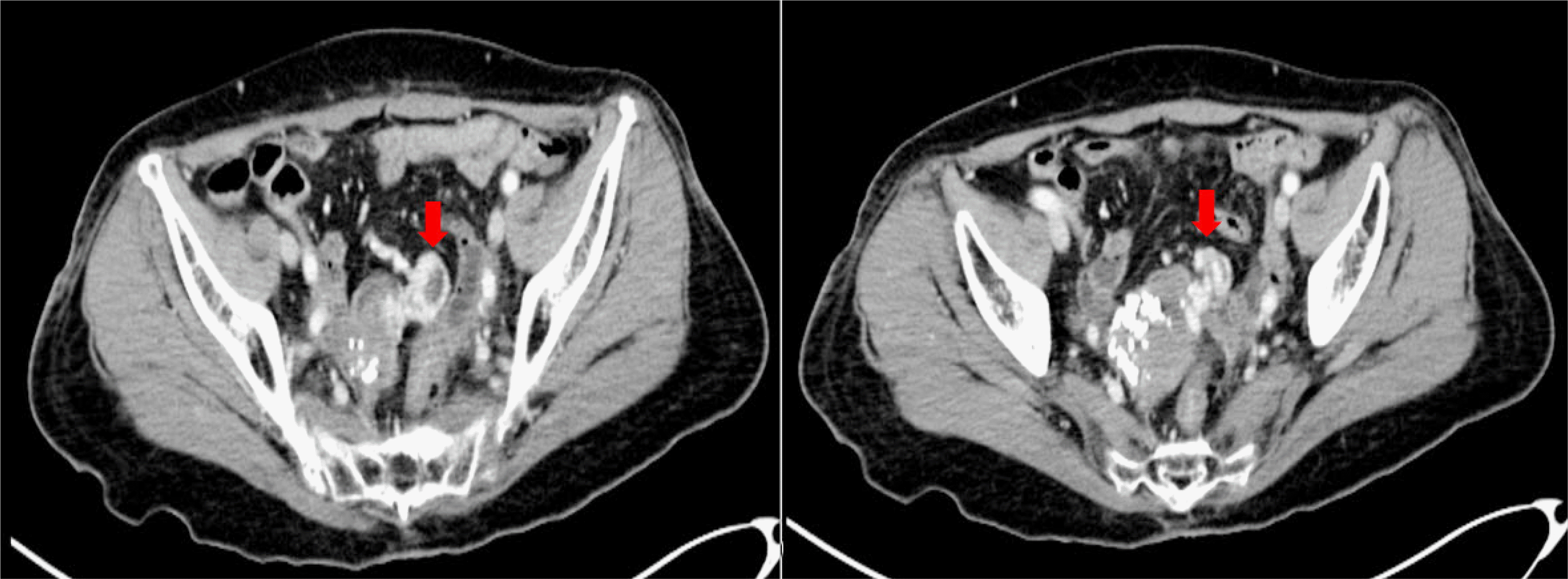
 | Fig. 3.Abdominal computed tomography revealed venous dilatation on the ileum (red arrows) adjacent to the uterus. |
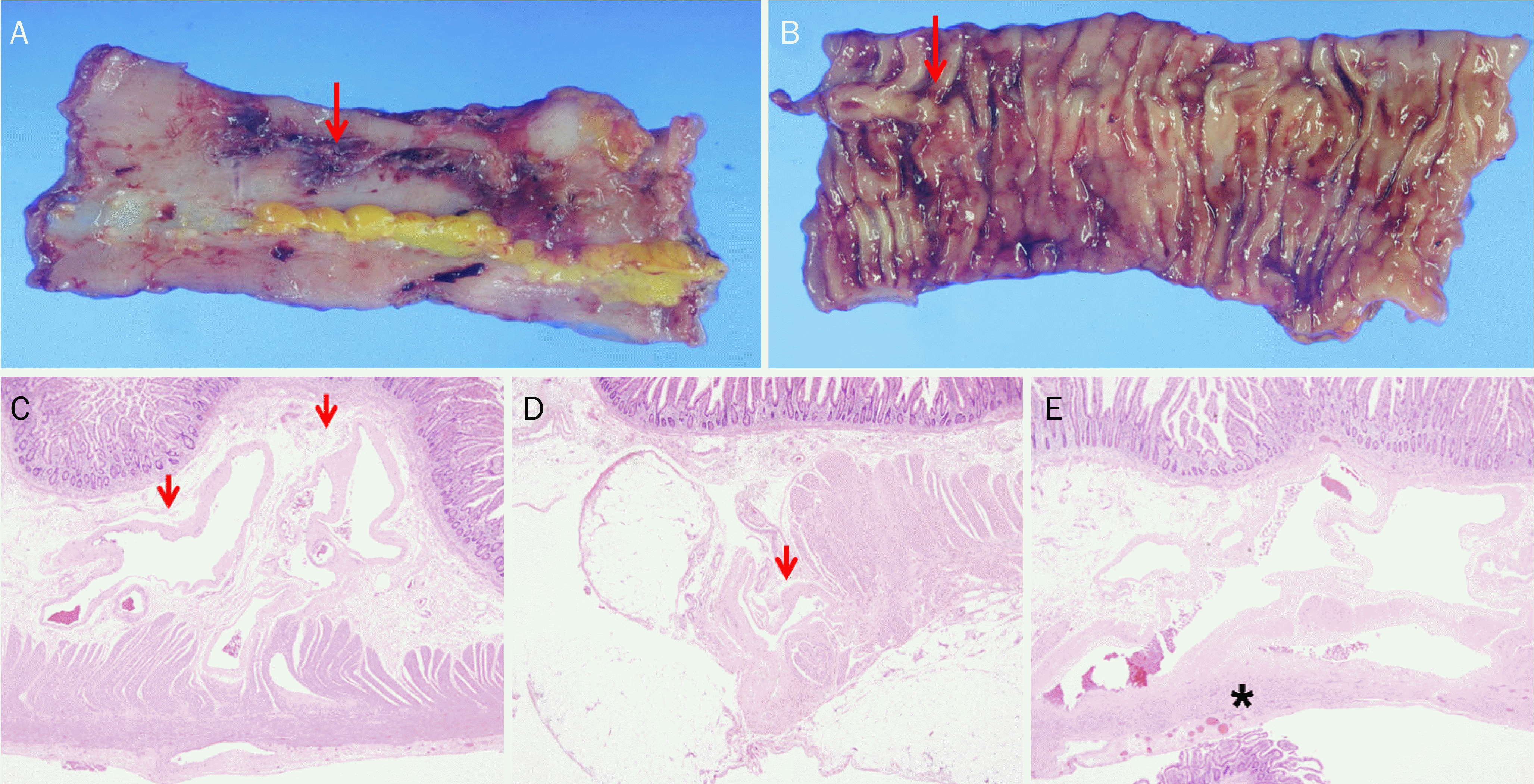
 | Fig. 4.Histopathology findings. (A, B) Gross findings of the resected ileum showed a well-defined tortuous, engorged vascular structure (red arrows) on the serosal (A) and mucosal (B) surface. (C, D) On a microscopic examination, submucosa (C, H&E, ×40) and subserosa (D, H&E, ×40) showed irregularly dilated venous structures (red arrows). (E) Fibrous bands (star) around the dilated tortuous varices were observed on the subserosa (H&E, ×40). |


 PDF
PDF ePub
ePub Citation
 Print
Print


 XML Download