

 PDF
PDF ePub
ePub Citation
 Print
Print


INTRODUCTION
Papillary thyroid carcinoma (PTC) is the most common malignant neoplasm of the thyroid gland. It may have a multifocal presentation in 18%–22% of all cases (1). However, the presence of 2 distinctive foci of different histological types of primary, well differentiated thyroid carcinoma in the same thyroid is a rare condition with only few reported cases (234). Moreover, a number of situations such as Hashimoto's thyroiditis (HT) and primary hyperparathyroidism (PHPT) were reported to be associated with well differentiated thyroid cancer, especially papillary cancer (56).
The current paper presents a female patient that underwent parathyroidectomy and thyroidectomy due to primary hyperparathyroidism and a suspicious thyroid nodule. The final pathology revealed not only a parathyroid adenoma (PTA) but 2 different, side by side, thyroid primary malignancies: follicular and papillary carcinomas, both in the same lobe, and in the background features compatible with HT. To the best of our knowledge, this is the first report of these thyroid and parathyroid pathologies occurring simultaneously. In addition, the possibility of interplay between 4 pathologies is discussed.
CASE REPORT
A 53-year-old healthy female was referred to our clinic having a growing central neck mass, accompanied by a disturbing pressure sensation while lying in the supine position. Ultrasonography of the neck revealed a non-homogenous thyroid texture with few nodules. The largest nodule was in the isthmus, 2.5 cm in its largest dimension. Adjacent to the right inferior thyroid lobe was demonstrated a 0.7 cm, hypoechoic nodule, suspected as a PTA. There were no suspicious lymph nodes signed. The largest thyroid nodule was aspired and the cytological results were compatible with follicular lesion with Hürthle cells (Bethesda IV). Calcium blood levels were at the height of the normal range (9.76 mg/dL), serum parathyroid hormone (PTH) and 24 hours urine calcium were 103 pg/mL and 278 mg/day, respectively (normal values: serum PTH up to 70 pg/mL and 24 hours urine calcium, 100–250 mg/day). A technetium-99m methoxyisobutylisonitrile imaging (99mTc-MIBI) suggested an atypical location for an adenoma, near the isthmus and to the left (Fig. 1).
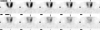 | Fig. 1
99mTc-MIBI scan. A suspected adenoma was located in vicinity to the inferior-medial left thyroid gland (arrows).
99mTc-MIBI = technetium-99m methoxyisobutylisonitrile imaging.
|
The patient was scheduled for a thyroidectomy and parathyroidectomy. During operation, the right thyroid lobe and isthmus were excised and sent for a frozen section examination. This study revealed a 2-cm follicular lesion with Hürthle cell features and another nodule having benign features although reported as having an undetermined diagnosis. Since a PTA was not found near the isthmus, within the right thyroid lobe or in the right neck, the left neck was explored as well. The few suspected specimens that were sent for a frozen section examination were all negative for a PTA, one reported as a large parathyroid gland. At this point the surgeon decided to complete a total thyroidectomy by excision of the left thyroid lobe. The considerations that guided this decision were having the 2 suspected lesions in the right lobe that may finally necessitate a total thyroidectomy and possibly having an intrathyroidal PTA. It is of importance to note that prior to surgery such a circumstance of having an unclear but suspicious frozen section report was discussed and consented with and by the patient for total thyroidectomy, being reluctant to have a second operation. The procedure, postoperative period and recovery were eventless. The intra-operative PTH blood levels dropped to normal (18 pg/mL) after the excision of the left thyroid lobe, and PTH, calcium follow-up measurements were normal.
1. Pathology
A follicular tumor with a maximal diameter of 2 cm was found within the right lobe of the thyroid gland. It was surrounded by a thick capsule composed of cells with abundant pinkish cytoplasm showing obvious capsular invasion, i.e, compatible with Hürthle cell carcinoma (Fig. 2). A second focus was demonstrated in the same lobe with a maximal diameter of 0.7 cm, with a margin of normal tissue separating both lesions. The cells of this tumor showed nuclear features of papillary carcinoma in the form of crowded ovoid nuclei with ground glass features, grooved nuclei and nuclear pseudo inclusions partly arranged in follicular fashion (follicular variant of papillary carcinoma) (Fig. 3).
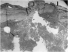 | Fig. 2Hürthle cell carcinoma showing invasion of tumor through the thick capsule at 2 foci highlighted by the black arrows (hematoxylin and eosin ×20).
|
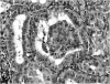 | Fig. 3Close-up view of the papillary carcinoma demonstrating a single papilla at the center of the field and the characteristic cytological features of papillary carcinoma (nuclear crowding, nuclear groves, and chromatin clearing) (hematoxylin and eosin ×200).
|
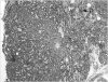
The non tumoral thyroid parenchyma demonstrated lymphoplasmocytic infiltration, follicular infiltration and destruction by lymphocytes as well as oncocytic changes in thyroid epithelium — features of HT. This was seen in the entire left lobe as-well, although with no malignancy or other focal pathology (Fig. 4).
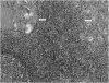 | Fig. 4Close up view of the non-tumoral thyroid parenchyma showing germinal center formation at the lower left, dense lymphoplasmocytic infiltration and small atrophic follicles with large oncocytic cytoplasm (arrows) compatible with Hashimoto's thyroiditis. (hematoxylin and eosin ×100).
|
A specimen of a parathyroid gland weighting 700 mg and a maximal diameter of 8 mm, free of adipocytes was compatible with PTA (Fig. 5)
 | Fig. 5Histologic picture of parathyroid gland demonstrating markedly increased cellularity and almost complete absence of adipocytes. Compatible with parathyroid adenoma (hematoxylin and eosin ×40).
|
It is noteworthy to mention that the adenoma was not clearly demonstrated by the US study. The 7-mm lesion that was described in relation to the right thyroid lobe and suspected to be an adenoma was in fact the papillary carcinoma. The 99mTc-MIBI scan suggested an atypical location for an adenoma, near the isthmus and to its left. Nevertheless, the adenoma was found adjacent to and medial to the left thyroid inferior pole.
DISCUSSION
Thyroid cancer accounts for approximately 2.5% of all malignancies and its incidence has been steadily increasing over the past 2 decades. Most thyroid carcinomas are well differentiated tumors of follicular cell origin. These lesions are histologically defined as papillary, follicular and Hürthle cell carcinomas. According to the World Health Organization classification, Hürthle cell tumor is a subtype of follicular cell neoplasm characterized by more aggressive clinical behavior and worse prognosis (7). Papillary carcinoma may be multifocal, representing either a de novo multicentric tumor or intraglandular metastasis. This feature is not a common characteristic of Hürthle cell carcinoma (8).
The current case report describes 2 coexisting, distinct and different histological types of primary thyroid carcinoma: papillary and Hürthle cell carcinoma in the same thyroid lobe. The incidence of 2 different histological types of thyroid cancer in the same thyroid is extremely rare and was described only in few case reports. Isildak et al. (2) described a case of a 72-year-old woman with a papillary thyroid microcarcinoma in one lobe and Hürthle cell neoplasm in the other. The remainder of the thyroid showed a typical pattern of colloidal goiter. Few other case reports presented collision tumors of the thyroid. The term “collision tumors” refers to the coexistence of 2 histologically distinct and morphologically independent malignant tumors within the same mass. Collision tumors can result of either 2 distinct tumors arising separately and growing one against the other or, one tumor that has arisen from the other. Two studies described collision tumors of papillary and Hürthle cell thyroid carcinoma (34).
The coexistence of papillary and Hürthle cell carcinoma in the same thyroid may either be anecdotal or may point at an association between the 2, questioning the assumption that Hürthle cell tumors are follicular tumor variants. At the genetic level, the rearranged during transfection (RET)/PTC oncogene which is the most frequent genetic alteration in PTC was shown to be common to both papillary carcinomas and Hürthle cell neoplasms (9).
The presented patient had 2 adjunctive conditions that were proposed to be associated with thyroid cancer, PHPT and Hashimoto. The incidence of having PHPT and a thyroid malignancy in the same patient was reported as 2%–15%, higher than expected in comparison to the general population, suggesting that patients with PHPT should be regarded as extra-suspicious for papillary thyroid cancer (6). The association of HT and thyroid cancer was attributed to the chronic inflammation process itself. Indeed, HT patients having elevated levels of anti-thyroglobulin antibody were reported to have more thyroid cancer. In addition, the enhanced expression of RET, RAS, and ERK proteins was found among the metaplastic oxyphil cells in HT and in PTC suggesting a possibility of a molecular link between HT and the progression of papillary thyroid cancer (510).
In conclusion, this case report demonstrated a unique combination of thyroid disorders. Although it may be an anecdote, these co-existing pathologies should intrigue us to look for interplay. Since all patients with PHPT would undergo an ultrasound, thyroid cytology should be addressed and attended cautiously and vice versa, serum PTH level should be measured for all patients undergoing thyroidectomy, looking for PHPT. Patients with Hashimoto and hyperparathyroidism having thyroid nodules should have a more cautious routine follow-up including cytology.
 XML Download