

 PDF
PDF ePub
ePub Citation
 Print
Print


INTRODUCTION
Walnut (WN) allergies, which can be fatal or near-fatal, account for a large proportion of total cases of tree nut allergies that affect children.12 Approximately 1% of the general population3 is allergic to nuts including WNs, although a trend of increasing incidence has been observed. WN imports to Korea have tripled in the last 10 years,4 and the consumption of WNs has become more widespread. There has been an increase in WN allergies among young children,56 and WNs are currently the third most common food to induce anaphylaxis and the fourth most common food to induce food allergy among Korean children.27 Additionally, regional differences have been found among young children in WN antigenicity patterns and severe systemic reactions after WN consumption.78
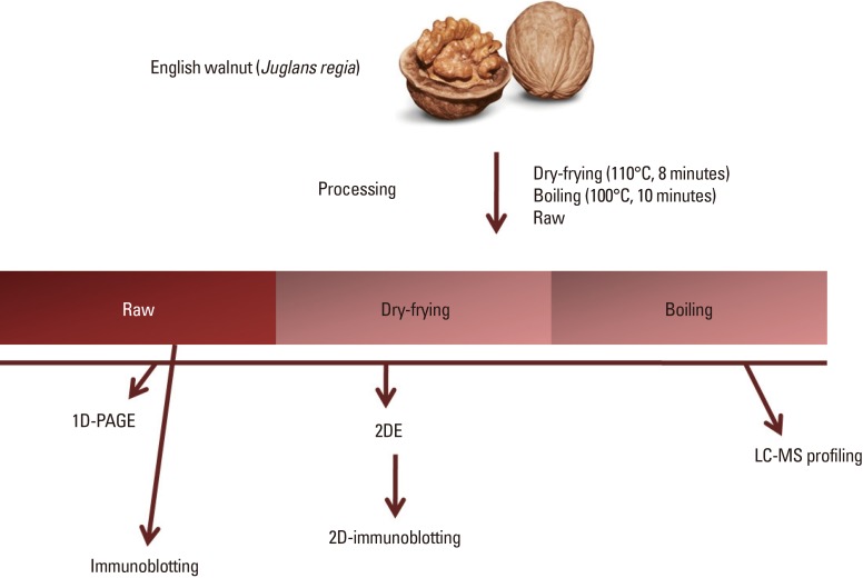
WNs, particularly English walnuts (Juglans regia), frequently undergo some form of thermal processing prior to consumption. Food processing methods have the potential to modify food proteins, which can alter food antigenicity.9 Moreover, some treatments such as dry-roasting can reduce immunoglobulin (Ig) E-binding capacity in vitro, measured via immunoblotting analysis.910 Pressure11 and thermal treatments910 have been used to diminish the cross-linking capacity of IgE to WN effector cells. However, previous studies which have focused on the decrease in allergenicity of WNs have employed thermal processing methods not used in domestic kitchens and have been performed using adult sera. The way Koreans cook is also different from Western countries. In Korea, WNs are mostly consumed raw, or eaten with rice. Often, WNs are boiled for 5 minutes in water and stir-fried with sugar, soy sauce and other seasoning for 10 minutes. Meanwhile, liquid chromatography tandem mass spectrometry (liquid chromatography-tandem mass spectropmetry [LC-MS/MS])12 can be used to assess changes in individual proteins or amino acid residues in a protein sequence, given the relative changes in key proteins as a result of thermal processing. Researchers have identified 5 WN allergens to date, namely, Jug r 1 (2S albumin), Jug r 2 (7S globulin), Jug r 3 (lipid transfer protein [LTP]), Jug r 4 (11S globulin), and Jug r 5 (profilin).
In the present study, we aimed to investigate the immunological characteristics of young Korean children with WN allergies. In addition, we identified the usual methods of cooking WNs in order to investigate changes in WN antigenicity caused by cooking. Furthermore, we used LC-MS/MS analysis to evaluate the antigenic changes in proteins from raw, dry-fried, and boiled WNs.
Go to : 
MATERIALS AND METHODS
Patients and sera
Between January 2011 and January 2016, we identified all children referred to Ajou University Hospital (Suwon, Korea) for the investigation of potential WN allergic reactions and enrolled patients under the age of 13 years who had WN serum-specific IgE (sIgE) concentrations, obtained via ImmunoCAP sIgE assays (Thermo Fisher Scientific, Waltham, MA, USA), of >0.35 kU/L (1.35–100.0 kU/L). The sera of the participants were stored at −20℃ until further testing. The clinical characteristics of the participants were collected from medical records and via telephone interviews with parents. Children with a clinical WN allergy were defined as those participants with a history of anaphylaxis and repeated systemic adverse reactions within 2 hours of isolated WN exposure. Food challenges were not performed for the majority of participants as parents frequently refused to provide consent. In addition, sera from 2 atopic patients who were allergic to inhalant but not food allergens were used as a control for immunoblotting. The study protocol was approved by the Institutional Review Board of Ajou University Medical Center (MED-KSP-12-381), and informed consent was obtained from the parents of all participants.
WN preparation and processing
Whole natural English WNs (Juglans regia) (Atlas World Food & Agriculture, Inc., Visalia, CA, USA) purchased from a local grocery store were used to obtain the protein extracts. The WNs were either dry-fried at 110℃ for 8 minutes in a frying pan, boiled in water at 100℃ for 10 minutes or left uncooked. The WNs were then ground in a blender until a smooth paste and defatted with cold (4℃) petroleum ether (1:1 w/v) stirred constantly for 1 hour, and then filtered through filter paper. The defatting procedure was repeated until the filtrate became clear. The defatted paste was air dried completely at ambient temperature, added to phosphate-buffered saline (PBS, pH 7.4; 1:10 w/v), and stirred for 48 hours at 4℃. The extracts were centrifuged twice at 14,000×g for 30 minutes at 4℃. The supernatants were then filtered and dialyzed against several changes of distilled water for 48 hours at 4℃. The extracts were centrifuged once again, lyophilized, and stored at -20℃ until use. Protein concentrations were then determined via a Bradford assay (Bio-Rad, Hercules, CA, USA) using a microplate reader (Bio-Rad).
One-dimensional polyacrylamide gel electrophoresis (PAGE) and immunoblotting analysis
The extracts were resuspended in PBS, and the protein concentration of each was measured using a Bradford assay. Equal concentrations of the samples were mixed with a loading buffer (60 mM Tris HCl, 25% glycerol, 2% sodium dodecyl sulfate, 14.4 mM 2-mercaptoethanol and 0.1% bromophenol blue) and heated for 10 minutes in a heating block at 100℃. The samples were then analyzed using sodium dodecyl sulfate (SDS)-PAGE, following which the gels were either stained with Coomassie brilliant blue or used for immunoblotting. After gel electrophoresis, the raw WN protein extract was transferred to a nitrocellulose membrane (Millipore Co., Bedford, MA, USA) and blocked in 3% bovine serum albumin (BSA) for 2 hours, washed 3 times in PBS-Tween, and placed in overnight incubation with clinically WN-allergic individual human and control serum (both 1:10 diluted with BSA) at 4℃. After washing, samples were conjugated with alkaline phosphatase-conjugated goat anti-human IgE (Sigma-Aldrich Co., LLC, St. Louis, MO, USA) diluted to 1:1,000. The membrane was then developed using SIGMA-FAST™ 5-bromo-4-chloro-3-indolyl phosphate/nitro blue tetrazolium (Sigma-Aldrich Co., LLC) and washed, and the development was terminated using water. Immunoblotting images were then analyzed using ImageJ software (National Institutes of Health, Rockville, MD, USA).
Two-dimensional electrophoresis (2DE) and 2D-immunoblotting analysis
Protein extracts from raw, dry-fried and boiled WNs were diluted with 50 and 300 µg/mL rehydration buffer (8 M urea, 2% 3-[(3-cholamidopropyl) dimethylammonio]-1-propanesulfonate, 3–10 NL 0.5% immobilized pH gradient [IPG] buffer, 0.002% bromophenol blue, and 50 mM dithiothreitol). The first-dimension separation was carried out on an Ettan IPGphor system (GE Healthcare, Chicago, IL, USA) in accordance with the manufacturer's instructions. The samples were loaded on 7- and 13- cm IPG strips, with pH 3–10 NL (GE Healthcare). Second-dimension separation was performed on 10% and 12% acrylamide gels, and colloidal Coomassie brilliant blue staining or electroblotting used for visualization. After electroblotting, the membranes were blocked using a similar procedure to that described above and incubated with 25 mL of the serum pool from WN sensitized subjects at a dilution of 1:3. Protein spots were visualized and analyzed using Typhoon 7000 FLA series and ImageMaster 2D Platinum 7.0 software (GE Healthcare).
LC-MS/MS for peptide analysis
Nano-LC-MS/MS was performed using EASY n-LC™ and LTQ Orbitrap XL™ mass spectrometers (Thermo Fisher Scientific), which were equipped with a nanoelectrospray source. The raw, dry-fried, and boiled WN extracts were separated on a C18 nanobore column (150.0×0.1-mm, 3-µm pore size; Agilent, Santa Clara, CA, USA). Mobile phase A liquid chromatography separation consisted of 0.1% formic acid and 3% acetonitrile in deionized water, and mobile phase B separation of 0.1% formic acid in acetonitrile. The chromatography gradient was designed for a linear increase from 5% B to 30% B in 40 minutes, 30% B to 60% B in 4 minutes, 95% B in 4 minutes, and 3% B in 6 minutes. The flow rate was maintained at 1,500 nL/minutes.
The mass spectra were acquired using data-dependent acquisition with a full mass scan (350–1,200 m/z), followed by 10 tandem mass spectrometry (MS/MS) scans. For MS1 full scans, the orbitrap resolution and automatic gain control were 15,000 and 2×10⁵, respectively; for MS/MS the automatic gain control in the LTQ was 1×10⁴ (Fig. 1).
Database search
The mascot algorithm (Matrix Science, Boston, MA, USA) was used to identify peptide sequences present in a protein sequence database. The database search criteria were: 1) taxonomy, fagales (National Center for Biotechnology Information non-redundant database, downloaded on Aug 4, 2016); 2) fixed modification, carbamidomethylation of cysteine residues, variable modification, oxidization of methionine residues; 3) maximum allowed missed cleavage of 2; 4) MS tolerance of 10 ppm; and 5) MS/MS tolerance of 0.8 Da. The peptides were filtered with a significance threshold of P<0.05.
Go to :
RESULTS
Participants and sera
A total of 20 participants who had sensitized to WNs (WN-sIgE>0.35 kU/L) were enrolled in the study. The median age of the participants was 36 months, with a range of 8–132 months. The study group was composed mostly of boys (75.0%, 15/20). Of the participants, 12 (60%) who presented with immediate hypersensitivity on isolated exposure to WNs were allocated to the clinical WN-allergic group. Based on the diagnostic criteria of the World Allergy Organization,13 3 participants had a history of anaphylaxis. Nine participants presented with repeated generalized skin rashes within 2 hours after WN exposure. In contrast, among all the sensitized participants, only 1 had no hypersensitivity reactions and 7 were too young to consume WNs due to the potential choking hazard (Table 1).
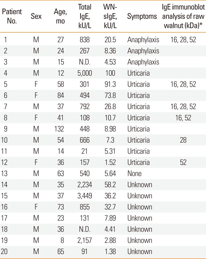
Table 1
Characteristics of patients with a history of walnut ingestion
![]()
Comparison of the protein fractions of raw, dry-fried and boiled WNs via SDS-PAGE and 2DE
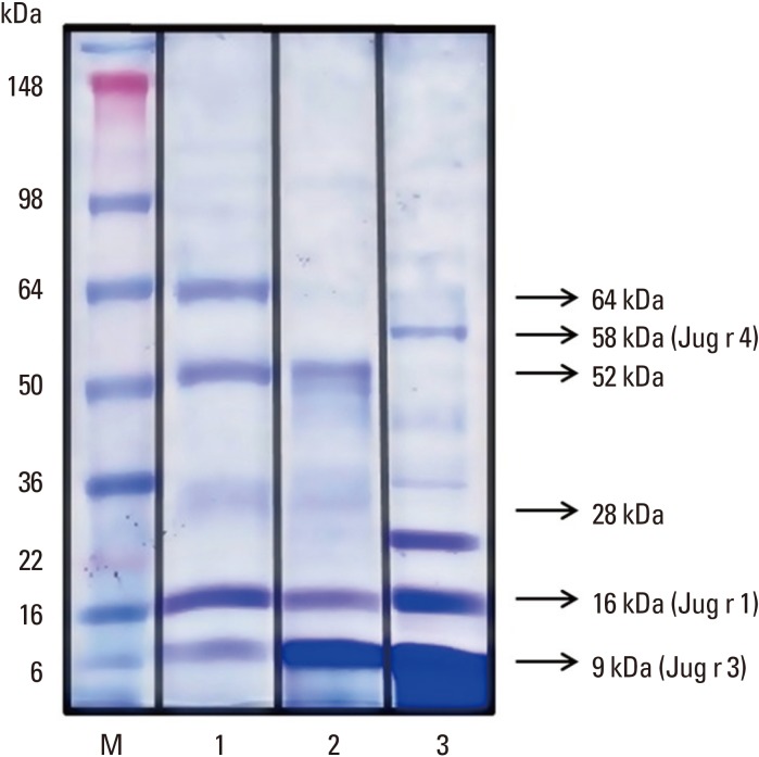
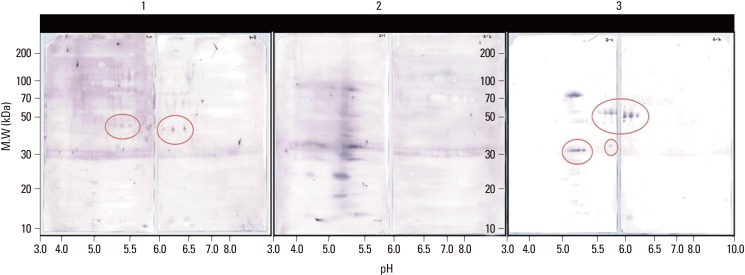
The protein fractions of dry-fried (3.4 mg/mL) and boiled (17.0 mg/mL) WN extracts were compared with those of raw (4.4 mg/mL) WNs using SDS-PAGE. The protein bands corresponding to raw WNs were 9, 16, 28, 52, 58, and 64 kDa. The patterns of each WN protein differed depending on the cooking method. The intensity of the 9-kDa (likely Jug r 3) and 16-kDa (likely Jug r 1) protein bands were enhanced by boiling. In contrast, the intensity of the 52- and 64-kDa protein bands almost completely disappeared with boiling. In addition, the intensity of the 9-kDa protein band was enhanced, whereas that of the 16-kDa protein band was reduced, by dry-frying. However, the intensity of the 28-kDa protein band was unchanged and a 58-kDa protein band was newly presented with boiling. Notably, the intensity of the protein band corresponding to Jug r 3 was enhanced by all treatments (Fig. 2). When we compared the 2DE protein profiles of raw, dry-fried and boiled WNs, we found that most protein spots dispersed after dry-frying. However, protein spots both dissolved and appeared spontaneously with boiling (Fig. 3).

IgE reactivity of each WN extract based on immunoblotting analysis
An immunoblotting analysis was conducted to examine IgE reactivity toward raw WNs using serum samples from the 6 patients with a clinical WN allergy (Patient Nos. 1, 5, 7, 8, 10, and 12; Table 1). The serum samples of 5 out of 6 participants (83.3%) reacted with the 52-kDa protein bands, and those of 4 out of 6 participants (66.7%) reacted with the 16- (likely Jug r 1) and 28-kDa protein bands, respectively, whereas none of the participants had reactions to the 9-kDa protein band, which we suggest was Jug r 3 (Table 1 and Fig. 4).

Meanwhile, to investigate the cooked WN extracts with IgE-binding capacity, a 2D immunoblotting analysis was performed using the pooled sera of the WN-sensitized patients (Patient Nos. 1–20; Table 1). The results confirmed the presence of different binding patterns among children who consumed cooked WNs (Fig. 5).
Protein profiles of differently processed WN extracts
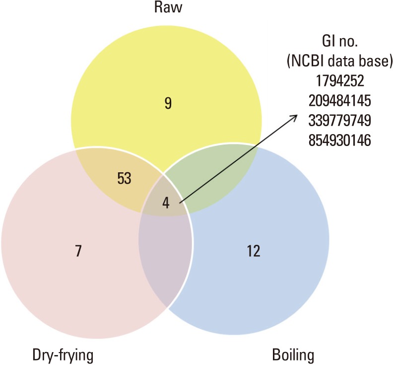
LC-MS/MS profiling of raw, dry-fried and boiled WN extracts revealed that 66 IgE-binding protein spots were observed in raw WNs; of these, 57 were also observed in dry-fried WNs. In contrast, only 4 IgE-binding protein spots were found in boiled WNs (Fig. 6). The 4 proteins that remained stable after all forms of preparation had high identification scores based on the J. regia database and included the albumin seed storage protein (Jug r 1), nsLTP (Jug r 3), glyceraldehyde-3-phosphate dehydrogenase (G3PD) C subunit and superoxide dismutase (SOD). In addition, 7 proteins were newly observed in dry-fried WNs and 12 proteins that appeared spontaneously in boiled WNs included vicilin seed storage protein (Jug n 2) and globulin seed storage protein (Jug r 4) (Table 2).
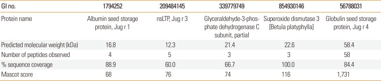
 | Fig. 6Overview of the number of protein bands observed in processed walnut extracts via the proteomic approach. Out of the 66 proteins present in raw WNs, only 4 were observed in boiled WNs. GI, GenInfo; NCBI, National Center for Biotechnology Information non-redundant database.
|
Table 2
Identification of differentially expressed proteins in processed walnut by LC-MS/MS
![]()
Go to :
DISCUSSION
Studies on antigenic changes in WNs due to heat treatments have been reported by a few institutions. The protein structure of Jug r 1 is known to be relatively stable at approximately 90℃, whereas Jug r 2 and Jug r 4 do not show antigenic changes when boiled for 5-10 minutes and are able to maintain their antigenicity when deep fried or microwaved at approximately 190℃.1415 Nevertheless, another study reported that Jug r 2 and Jug r 4 lose their protein bands when roasted after boiling in water.11 However, the aforementioned studies were not conducted on patients, and no studies have shown which allergenic components of cooked WNs cause serious reactions when consumed by children. In Korea, WNs are eaten with rice and usually boiled and stir-fried with seasoning. Accordingly, in this study, we investigated major WN allergens identified in Korean children and changes in the antigenicity of cooked WNs compared to raw WNs. Our aim was to identify the target component allergens in WNs in order to provide safer dietary advice to allergic children.
We analyzed antigenic changes in WNs cooked according to usual Korean methods (dry-frying and boiling) using SDS-PAGE, 2DE and LC-MS profiling. Our investigation of the effects of different cooking methods showed that 57 out of the 66 proteins found in untreated WNs were also identified in the dry-fried WNs, whereas only 4 out of the 66 proteins were identified in the boiled WNs. This shows that boiling resulted in lower antigenicity than dry-frying. Previous studies of component allergens using recombinant allergen technology have found that storage proteins and LTP allergens in nuts are relatively stable to heat, which was consistent with the findings of the present study. After boiling, we were able to identify a 9-kDa protein (LTP), a 16-kDa protein (2S albumin), 21- and 23-kDa proteins (G3PD and SOD) remained unchanged. Also, Jug r 4, a member of the 11S globulin seed storage protein family, has been characterized by its distinct antigenicity, which newly appeared after boiling; this finding was consistent with our results from SDS-PAGE. The 2 allergens G3PD and SOD may have originated from birch or oak pollen, which acted as the primary sensitizer or may have cross-sensitized due to homology with WN allergens. Additional clinical studies on this topic are necessary.
When we analyzed individual immunoblots of IgE-binding proteins using blood serum from the 6 children with clinical WN allergies, our results indicated that the 16-, 28- and 52-kDa proteins, rather than the 9-kDa protein (LTP) of WN, are more likely to contribute to the adverse reactions among Korean children. In addition, in this study, the 52-kDa band almost completely disappeared with boiling. These findings suggest that when patients solely sensitized to 52-kDa protein, there is a low probability of consumption of cooked WNs prepared according to common Korean recipes causing serious allergic reactions and that the 16-kDa protein band (Jug r 1, storage protein) may be the most potent allergen among Korean children. Also, Jug r 1 was, as found in previous studies, generally stable under both cooking methods. Thus, clinicians should highlight this finding when educating their patients on the antigenicity of cooked WNs.
This study has several limitations. First, changes in antigenicity based on digestive enzymes were not considered. Thus, the study did not completely replicate in vivo conditions. Secondly and most importantly, although an in vitro study measuring IgE reactivity can give information on allergenicity, clinical studies using food challenges are considered to provide the highest level of evidence. Further research including prick-to-prick and food challenge tests, which can document changes in the eliciting cooked WN dose, are needed to determine the hypoallergenicity of processed WNs. Thirdly, the analysis had a limitation of including a small number of patients, thus being a preliminary result.
Nevertheless, this study has several strengths. First, we have demonstrated that Korean children are highly likely to be sensitized to Jug r 1. We have also found that LTP is not a major allergen among this population. This result differs from those of previous studies mainly performed in Europe, and indicates which allergens need more focus in Korea. Secondly, when investigating antigenicity of the various WN proteins, we found that the storage protein Jug r 1 was stable under casual cooking methods. Previous studies that have focused on changes to the allergenicity of WNs have used cooking processes that do not replicate those used in everyday life. Although we found that the allergenicity of certain components of WN diminished, clearly cooked WNs must not be given to Korean children with WN allergies. Thirdly, the findings of this study support existing ones that the allergenic components of WNs have varying antigenicity depending on the cooking method. Therefore, the allergenic components of WNs identified via diagnostic tests of the allergic participants in this study could potentially be used to justify different dietary restrictions on WNs cooked in different ways.
In conclusion, we suggest that further studies are needed with a focus on the storage protein Jug r 1 in young Korean children for stable oral food challenges in high risk patients and the development of low-allergen WNs that can be cooked at home. In the future, these low-allergen WNs could be used for desensitization therapy of patients who are allergic to WNs. LTP allergen (Jug r 3) appears to be one of the candidate allergens causing severe reactions in patients with LTP syndrome,1617 but not in this study. In order to draw more definite conclusions on LTP, further studies on older participants are needed. Additionally, since plant-derived panallergens, such as SOD and G3PD may be problematic for children allergic to nuts, future studies on these allergens, which have been neglected to date, are also necessary.
Go to :
 XML Download