

 PDF
PDF ePub
ePub Citation
 Print
Print


Introduction
Periodontal diagnosis requires a thorough clinical examination, involving a consideration of the patient's signs, symptoms, medical history, and dental history. Radiographic evaluation, in turn, plays a decisive role in confirming and establishing diagnoses by providing information on the type and severity of damage to the alveolar bone.1 For this purpose, a series of conventional radiographic techniques have been used, the most common of which are bitewing, periapical, and panoramic radiographs.2
The establishment of sensitive radiographic techniques and 3-dimensional methods for assessing dentoalveolar structures led to the development of new techniques such as digital imaging, which enabled the manipulation of contrast and density levels using specialized software. In addition, a smaller dose of radiation is required to sensitize digital sensors than is needed for conventional films. Nevertheless, digital radiographs do not overcome certain limitations of conventional 2-dimensional imaging techniques, such as the risk of overestimating or underestimating the amount of alveolar bone loss.3456
The advent of cone-beam computed tomography (CBCT), in turn, offered solutions to some of the abovementioned limitations. CBCT involves a conically shaped X-ray beam, directed at a region of interest, which sensitizes a 2-dimensional array of image detectors.7 This modality has brought a series of benefits to the field of diagnostic imaging, including the elimination of distortions and the ability to visualize structures in all 3 orthogonal planes.89 Although CBCT is a promising technique in the field of periodontal diagnosis, it is still more expensive than conventional techniques and involves a higher radiation dose.10
A few studies using CBCT for periodontal diagnosis have been conducted, and have shown potential benefits.11 However, little is known regarding comparisons between conventional and 3-dimensional imaging techniques in terms of diagnostic performance, precision, accuracy, and advantages and disadvantages in specific circumstances. Therefore, the objective of this study was to carry out a systematic review of studies in the literature comparing conventional imaging techniques and CBCT for the assessment of infrabony defects, furcation involvement, height of the alveolar bone crest, and the periodontal ligament space.
Materials and Methods
In this study, a systematic review design was adopted to compare the precision and accuracy of conventional imaging techniques and CBCT for infrabony defects, furcation involvement, height of the alveolar bone crest, and the periodontal ligament space.
The MEDLINE (via PubMed) and Embase databases were searched for English-language articles published through 2017 using the following search strategy developed for MEDLINE: ((CBCT OR cone beam computed tomography) AND (digital radiography OR digital radiology) AND (periodontal OR periodontitits) AND (diagnosis OR diagnostic accuracy)). The reference lists of articles considered for inclusion and the OpenGrey12 database were screened for relevant unpublished studies and papers not identified by the electronic search.
Original articles, systematic reviews, and case reports were considered for inclusion. Interventional studies were included if they reported clinical outcomes from adult patients with infrabony defects, furcation involvement, or marginal bone loss. Book chapters and conference abstracts were excluded from the study. To be considered eligible for inclusion, studies must have reported the advantages and disadvantages of visualizing some of the above periodontal conditions with more than 1 diagnostic imaging technique. Studies that did not compare CBCT findings with results obtained using other diagnostic modalities were not considered eligible for inclusion. The review text was structured in accordance with the PRISMA (Preferred Reporting Items for Systematic Reviews and Meta-Analysis) guidelines.13
Data extraction
Two independent reviewers with expertise in periodontal research screened the titles, abstracts, and full texts of the articles that were identified. When considered necessary, attempts to contact the authors were made. The following data points were extracted and recorded: year of publication, location of the study, characteristics of the groups or sample, methodological characteristics, outcome measures, conclusions, and source of funding.
Results
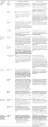
A total of 351 potentially eligible papers were screened. Of these, 328 were excluded after title, key word, and/or abstract assessment, yielding 23 papers that potentially met the inclusion criteria. Ten additional studies were excluded because their full texts did not present any direct comparisons between a conventional imaging technique and CBCT. Thus, 13 studies were ultimately identified as eligible for inclusion in this systematic review. The characteristics of the included studies are summarized in Table 1.
Comparison of clinical findings and CBCT data
Among the included publications, 4 discussed infrabony defects, 3 discussed the height of the alveolar bone crest, 3 discussed furcation involvement, 2 discussed the periodontal ligament space, and 1 discussed infrabony defects, furcation involvement, and the periodontal ligament space.
In 2 of the studies that analyzed infrabony defects, the findings were obtained from naturally occurring defects in human skulls,1415 while the other 2 evaluated simulated infrabony defects.1617
Of the studies that evaluated the height of the alveolar bone crest, 1 imaged bone defects in human skulls,18 another was an in vivo study that evaluated correlations between CBCT and direct surgical measurements after bone-replacement graft procedures,19 and the third analyzed images selected from a secondary database containing images of patients referred for periodontal evaluation.20
Two of the three studies that were included because they discussed furcation involvement utilized intraoperative findings as the gold standard,2122 while the third analyzed simulated lesions in 15 macerated pig mandibles.24
The 13th study was a human ex vivo study that used histological evaluations as the gold standard and confirmed close agreement between the histological findings and CBCT in terms of the identification of infrabony defects, furcation involvement, and the periodontal ligament space.23 Only 4 of the 13 publications were in vivo studies, and all of them used different scanning parameters, making it difficult to compare their findings directly.
Infrabony defects
The studies performed to evaluate artificially created infrabony defects, comparing measurements made using periapical radiographs and CBCT with control measurements obtained directly from dry skulls, reported that higher precision and accuracy were obtained using CBCT. The greater precision of CBCT measurements can be attributed to the advantage of identifying defects present in both the buccal and lingual aspects of the teeth. Such defects cannot be fully identified by conventional radiography.141617 As a result, all artificially created defects were detected with CBCT, whereas only 67% of the defects were identified in periapical radiographs.16 Studies have demonstrated that CBCT has the potential to be more accurate17 than conventional techniques in assessing artificially created periodontal defects.1524
Only 1 study evaluated the accuracy of CBCT in identifying clinical bone defects, such as fenestrations and dehiscences.18 The authors of that study compared observations made using tomographic images with direct observations of the defects in the skulls. The number of fenestrations detected by CBCT was 3 times higher than the number detected by direct visualization, thus leading to several false positive results (104 observed versus 32 actual fenestrations). In contrast, fewer dehiscences were found in the tomographic images than by direct visualization (43 observed versus 52 actual dehiscences).14 The authors suggest that the low accuracy could be attributed to certain natural aspects of clinical bone defects, which present with less well-defined margins than artificially created defects, or to limitations in the spatial resolution of the CBCT device used.
Another study comparing measurements of periodontal defects (fenestrations, dehiscences, and furcations) made using periapical radiographs, panoramic films, CT, and CBCT with the corresponding histological specimens showed that image quality (contrast, brightness, distortion, overlay, and clarity of structures) was superior using CBCT. CBCT scans showed smaller deviations in the dimensions of the periodontal defects than were obtained using the histological results.23
Height of the alveolar bone crest
CBCT was found to be more accurate than conventional radiographs for assessing alveolar crest height, because it led to less underestimation of horizontal bone loss.1820 One of the studies found that the mean alveolar bone crest was 0.23 mm higher in the CBCT images than the actual crest height, whereas the corresponding deviation was 1.17 mm in periapical radiographs.18 Similar results were also obtained by de Faria Vasconcelos et al. (2012),20 who evaluated 51 dental sites.
Nevertheless, the diagnostic accuracy of CBCT images achieved in the study by Mol and Balasundaram (2008)18 for horizontal bone loss in the anterior teeth was low. Both the underestimation of bone loss and the poor accuracy of measurements of crest height in the anterior maxilla could be attributed to the very thin cortical bone present in this region and the low sharpness of the images produced by the CBCT device.
One of the few in vivo studies comparing bone height measurements from digital intraoral radiographs and CBCT images to direct surgical measurements for the evaluation of regenerative treatment outcomes confirmed that CBCT yielded satisfactory results. In that study, digital IR and CBCT images were taken prior to initial bone grafting and after a 6-month follow-up. A total of 35 intrabony defects were analyzed. The results suggested that CBCT reliably detected graft resorption based on a comparison with direct surgical measurements.19
Furcation involvement
CBCT images were highly accurate (78–88%) in detecting artificially created furcation involvement in 15 macerated pig mandibles in the in vitro study conducted by Umetsubo et al. (2012). Furthermore, furcation defects were also identified more accurately in CBCT images than in intraoral digital radiography in the study by Vandenberghe et al. (2008).1524 Some in vivo studies also evaluated furcation involvement in tomographic images that were compared with direct intrasurgical assessments as the gold standard. The CBCT and surgical measurements agreed in 84% of cases, which is in accord with previous studies that found similar results.2122 In the consensus report of the AAP Regeneration Workshop, the use of 3-dimensional radiographic modalities was encouraged for the assessment of furcation defect treatment outcomes.25
Periodontal ligament space
Few studies have evaluated the accuracy of imaging techniques for evaluating the periodontal ligament space, and the existing studies have reported different results. Some studies have shown that CBCT was more accurate than conventional radiography in detecting marginal widening of the periodontal space.2326 For instance, Jervoe-Storm et al. (2010) found almost 100% accuracy in identifying periodontal ligament space widening on CBCT images.26 Ozmeric et al. (2008), in contrast, found that periapical radiography was superior to CBCT for periodontal spaces smaller than or equal to 200 micrometers.27 The authors of that study suggested that the observers' lack of experience with CBCT may have favored the results from 2-dimensional conventional images.
Discussion
Some CBCT parameters are known to lead to higher or lower spatial resolution. In the study of Leung et al. (2010),14 the authors reported that the low accuracy of CBCT images in identifying clinical bone defects was due to limitations in the spatial resolution of the CBCT device used. The smallest thickness of bone measured in the axial and coronal slices was 0.6 mm, which may explain the large number of false-positive fenestrations found in the maxilla, where the cortical bone is less dense. In addition, the aforementioned study also had image quality limitations. Image quality is also influenced by the scanning parameters of the CBCT device. In this context, lower milliamperage (mA) leads to lower contrast resolution.28 Misch et al.16 (2006) chose to use high milliamperage in their research (47.7 mA, 120 kVp, and 20 s) and obtained satisfactory results for identifying artificially created defects. In contrast, Leung et al.14 (2010) chose to use lower milliamperage (2 mA, 110 kVp, and 9.6 s) and obtained lower accuracy and sensitivity in their measurements and classifications.
In the study of Mol and Balasundaram (2008),18 bone loss detection was significantly better with CBCT than with conventional intraoral radiographs, but the diagnostic accuracy of both imaging modalities was low for the anterior teeth. These results could have been better, as the authors reported image quality limitations because of the obsolete CBCT device used in their study. Advances in spatial resolution and other parameters have been incorporated into most modern devices, improving the resolution and precision of tomographic images.22 Thus, further investigations with modern devices using optimal parameters can be recommended to address the feasibility of CBCT.
The studies performed of CBCT data for furcation involvement not only showed high accuracy in identifying different levels of furcation involvement, but also demonstrated that the CBCT images provided additional important information about the root morphology and residual attachment of maxillary molars, which is a significant advantage of CBCT over conventional pre-surgical clinical assessment methods.21 Furthermore, the differences reported in the studies of furcation involvement may have been due to different levels of expertise among the observers, who should be also trained to properly manipulate the CBCT software. In the study conducted by Umetsubo et al. (2012),24 for instance, moderate interobserver agreement was found (κ= 0.40–0.59), indicating a lack of reproducibility.
Another example of how CBCT parameters can interfere with the final diagnosis of various periodontal conditions was provided by the study of Jervoe-Storm et al. (2010).26 The authors found an accuracy of almost 100% in identifying periodontal ligament space widening, and that result was probably obtained due to the high resolution used for imaging, as reflected by the voxel size of 0.15 mm. This finding is in contrast with that of Ozmeric et al. (2008),27 who reported that periapical radiography was superior to CBCT for periodontal spaces smaller than or equal to 200 micrometers, using a voxel size of 0.7 mm.
A limitation of the articles reviewed herein is that most of them were based on in vitro experiments. Only 4 of the 13 articles were in vivo studies. Another limitation was variation in the levels of expertise of the observers who participated in the research. Observers should be very well trained in manipulating the CBCT software, but several authors reported that observers experienced difficulties in manipulating the CBCT software to visualize the different periodontal parameters.
The lack of studies that qualitatively or quantitatively compared conventional imaging techniques and CBCT was another limitation. The most recent in vitro/in vivo research found was from 2014.
CBCT is significantly more accurate and reliable than 2-dimensional conventional imaging techniques for assessing infrabony defects, furcation lesions, the height of the alveolar bone crest, and the periodontal ligament space. However, differences in imaging protocol parameters can affect the reproducibility and reliability of CBCT measurements.
 XML Download