

 PDF
PDF ePub
ePub Citation
 Print
Print


Radiopacity in the maxillary sinus can be observed in various conditions, such as in the presence of lesions in the maxillary sinus or as a sequela of maxillary sinus surgery.1 Herein, we report a rare case of osseous metaplasia with bone and fibrous tissue in the bilateral maxillary sinuses in a patient with no relevant history of surgery or trauma.
Case
A 57-year-old female patient was referred for evaluation of the maxilla and oral cavity. She had a 2-year history of occasional nasal pain and no history of medical problems, trauma, surgical treatment of the nose or maxillary sinus, or use of drugs related to bone metabolism. The clinical examination at admission did not reveal nasal obstruction, postnasal drip, foul odor, anosmia, or snoring.
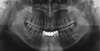
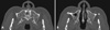
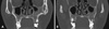
In panoramic radiographic images taken for a dental evaluation, both maxillary sinuses were indistinguishable and showed signs of radiopacity (Fig. 1). Computed tomography images showed bony tissue filling the maxillary sinuses, as well as signs of sinus mucosal hypertrophy (Figs. 2 and 3). Ossification was predominantly observed in the left maxillary sinus, and was detected to some degree in the right frontal sinus region, but not in the ethmoid region. To examine the bony tissue, an intraoral incisional biopsy of the left maxillary sinus was performed under local anesthesia. Biopsy analysis revealed trabecular bone fragments and fibrous tissue without cortical bone (Fig. 4). These histologic findings confirmed that the radiopaque lesion of the maxillary sinus was osseous metaplasia.12 The patient was advised to visit the hospital again if her symptoms of rhinitis recurred.
Discussion
Maxillary sinus ossification has been described in various conditions. Ossification can occur at the base of the maxillary sinus following maxillary sinus floor elevation for dental implant placement.3 Moreover, surgical treatments, such as radical curettage of the maxillary sinus, can induce a pattern of heterotopic ossification originating from the bone at the maxillary sinus wall.1 However, the patient in the present case had no history of trauma or surgical treatment of the maxillary sinus or surrounding tissue. Radiopacity of the maxillary sinus has also been observed in cases of tumorous lesions of the maxillary sinus, which can be confirmed by biopsy based on the pattern of growth and invasion of surrounding anatomical structures.4 Ossification in both maxillary sinuses should be differentiated from other lesions, such as antroliths and fungal disease of the maxillary sinus.5
Maxillary antroliths are masses formed by the gradual deposition of mineral salts. Calcium phosphate and calcium carbonate are endogenous minerals that can form antroliths.5 In this case, a dense and irregular, yet well-defined mass was radiographically identified in the maxillary sinus antrum. Antroliths must be included in the differential diagnosis of radiopacities found in or near the maxillary sinus region.6 They can be ruled out because antroliths form around a nidus or concentrated mucus and grow heterogeneously, with well-defined concentric layers extending from the maxillary sinus lateral wall. Aspergillus should be considered, as it is the most common pathogen involved in fungal sinus disease. It can cause complete or near-complete opacification of the sinus cavity, and is associated with thickening of the sinus mucosa.5 This point may be relevant in the differential diagnosis between Aspergillus infection and osseous metaplasia of the maxillary sinus. Other possible diagnoses include complex odontoma, mature cementoma, foreign bodies, and osteoma, which has the radiological appearance of a more or less homogeneous radiopaque lesion that is attached to the sinus wall with a small stem.4
Osseous metaplasia involves the bone in the maxillary sinus, including the Haversian system and bone marrow.2 When a biopsy is required, the oral vestibular approach under local anesthesia can be useful, as was performed in the present case. The cause of osseous metaplasia in the present case is unknown.
Osseous metaplasia of the maxillary sinus, which appears as heterotopic ossification in radiographic images, should be considered as an additional possibility in patients with no history of surgery or trauma to the maxillary sinus.

 XML Download