

 PDF
PDF ePub
ePub Citation
 Print
Print


Helicobacter pylori is the most important etiological agent of chronic gastritis and peptic ulcers and also increases the risk for gastric cancer [12]. Accumulating evidence has demonstrated that eradicating H. pylori in the stomach by administering oral antimicrobial agents results in the resolution of H. pylori-associated gastroduodenal diseases [34]. In addition, eradication of H. pylori also decreases the risk for gastric carcinogenesis [5]. Triple combination therapy, using two antibacterial antibiotics and a proton pump inhibitor, achieves a high eradication rate [6]; however, such combination therapy does not always successfully eradicate H. pylori.
Antibiotic chemotherapy occasionally produces side effects and fails to eliminate the bacterial infection [7]. The occurrence of strains resistant to antibiotics is expected to increase; thus, it is important to search for nonantibiotic substances to cure these infections [8].
Allium species contain an abundance of organo-sulfur compounds, volatile sulfur compounds, proteins, prostaglandins, fructan, vitamins, and polyphenols, and particularly, its sulfur and numerous phenolic compounds make them great of interest [9]. Allium hookeri which is a member of family Alliaceae subgenus Amerallium, is found in Ceylon, Greece, Yunnan, Southern China, Bhutan, Sri Lank, and India. These plants have been used by the locals to treat cough and cold, and heal burns and wounds [10]. Several studies have demonstrated antioxidant activity and anti-inflammatory effect of Allium hookeri [111213], however, there is still insufficient information available on the anti-helicobacter effect of Allium hookeri extract.
The goal of this study was to determine the anti-Helicobacter pylori activity and inhibition of Helicobacter pylori-induced inflammation effects of the Allium hookeri root extract (AHE).
Materials and Methods
Preparation of Allium hookeri extract
The dry mass of Allium hookeri was purchased from an Oriental Pharmacy (Iksan, Korea), was according to the standard as mentioned in Korean Pharmacopoeia and Korean Herbal Pharmacopoeia, which are the official compendia of standard. The procedure for preparing AHE was as follows. The air-dried mass of Allium hookeri (100 g) was cut into pieces and extracted twice with 70% (v/v) ethanol (three times as much as the weight of the dried plants) for 3 hours at 100℃. After filtration through a 400-mesh filter cloth, the filtrate was refiltered through filter paper (Whatman, No. 5) and concentrated on a rotary evaporator (N-2110, EYELA, Tokyo, Japan) and the concentrated filtrate was evaporated to dryness under vacuum with freezing dryer (FreeZone 2.5, Labconco, Kansas, USA). Finally, the solid residue was collected, placed in sealed bottles and stored at −20℃.
Determination of anti-bacterial activity
The in vitro anti-bacterial activities of AHE were determined by disk agar diffusion method [14]. Briefly, a total volume of 100 µL of H. pylori suspension (1×10⁸ colony forming units (CFUs)/mL) was spread onto Mueller Hinton agar plates (BBL) containing 10% sheep blood. Sterile paper disks (6 mm, BBL) were placed on the agar surface with 3 different concentrations of AHE extracts (12.5, 25 and 50 µg/mL) individually. DMSO was used as negative control and antibiotics amoxicillin (AMX, 0.05 mg/mL), clarithromycin (CLR, 0.05 mg/mL), metronidazole (MTZ, 0.8 mg/mL), and alliin (0.05 mg/mL) were used as positive control. After 72 h for incubation at 37℃ under the microaerophilic condition with humidity, the inhibition zone was determined in diameter.
Animals
5-week aged Specific pathogen-free (SPF) male C57BL/6 mice were procured from Samtako Co. (Osan, Korea). All animals were kept at the inspecting facility of Wonkwang University (Iksan, South Korea) for 1 week to allow acclimation before experimentation. Thereafter, they were kept in an isolated SPF barrier room with regulated temperature (23±1℃), humidity (50±5%) and light/dark cycle (12/12 h). The animals were fed a sterilized (2 M rad radiation) pellet diet (Purina, Seoul, Korea) and sterilized water ad libitum. All studies were performed in accordance with the Guide for Animal Experimentation of Wonkwang University and approved by the Institutional Animal Care and Use Committee of Wonkwang University. All efforts were made to minimize pain or discomfort to the animals used.
Bacterial inoculation
H. pylori SS1 (B0890, Helicobacter pylori Korean Type Culture Collection, HPKTCC) was incubated in brain-heart infusion broth containing 10% fetal bovine serum at 37℃ overnight under a micro-aerophilic atmosphere and allowed to grow to a density of ~2.0×10⁹ CFU/mL culture broth. Animals were inoculated 3 times at 3-day intervals by intragastric inoculation of 1.0×10⁹ CFU H. pylori suspended in 0.5 mL broth. The challenged animals were confirmed to be H. pylori-positive by the stool antigen analysis with SD Bioline H. pylori Ag kit (Standard Diagnostics, Inc, Yongin, Korea) as described previously [15].
In vivo study protocol
The inhibition effect of the AHE on H. pylori infection was investigated using a mouse model. Mice were divided into four groups: negative control (group I, n=10); H. pylori-infected, AHE-untreated animals (group II, n=10); and H. pylori-infected, AHE (25 mg/kg)-treated animals-treated animals (group III, n=10); and H. pylori-infected, AHE (50 mg/kg)-treated animals (group IV, n=10); and H. pylori-infected, AHE (100 mg/kg)-treated animals (group V, n=10). The AHE was administered orally daily at 25, 50 and 100 mg/kg/day dose during a 4 week treatment period.
At 4 weeks after H. pylori inoculation, all animals were sacrificed and their stomachs were dissected after ether euthanasia. Each stomach was opened along the greater curvature and washed with saline. Then, half of the glandular mucosa was scraped for detecting colonizing H. pylori, and the residual part was formalin-fixed and embedded in paraffin for histological observations. H. pylori colonization was confirmed by rapid urease tests, as described previously [16]. Mucosal damage was evaluated grossly and histologically according to previously described criteria [16].
Statistical analysis
Values for all parameters under study were recorded for each experimental unit, and the statistical analysis was performed using a general linear model. Values are reported as average ± standard deviation, when appropriate. Student's t-test was used for pair-wise comparisons. Incidence percentages (95% confidence intervals [CIs]) were calculated with the MiniTab statistical software program (Minitab, State College, PA, USA). A P-value <0.05 was considered significant.
Go to : 
Results
Yield of Allium hookeri extract
The extract yield of the Allium hookeri extract with 70% ethanol was 12.54%. We analyzed AHE composition by LC. The retention time of alliin in the specified LC condition was 9.667 min. The concentration of alliin in AHE was 710 µg/mL
Growth inhibition of H. pylori
We conducted the investigation of relative inhibitory potency of AHE against H. pylori SS1 growth, using the disk agar diffusion assay. AHE were tested at a maximum concentration of 100 µg/mL. For referencing, inhibitory effects of standard antimicrobial agents for treatment of H. pylori infection, with CLR and AMX at 50 µg/mL, and metronidazole (MTZ) at 800 µg/mL, and alliin at 50 µg/mL, were determined as well. To quantitative the inhibitory effect of H. pylori, the diameter of growth inhibition area was measured and expressed in millimeters and presented in Table 1. As shown in Table 1, tested agents showed a wide range of inhibitory effect against H. pylori growth with inhibition zone ranging from 0 to 20.2 mm. The antibiotics CLR, AMX and MTZ inhibited the bacterial growth with inhibition zone of 19.5, 15.6 and 7.6 mm, respectively. Alliin, an active ingredient of Allium hookeri, inhibited the bacterial growth with inhibition zone of 15.6 mm. AHE 100 µg/mL showed an inhibition zone value of 20.6 mm (Table 1).
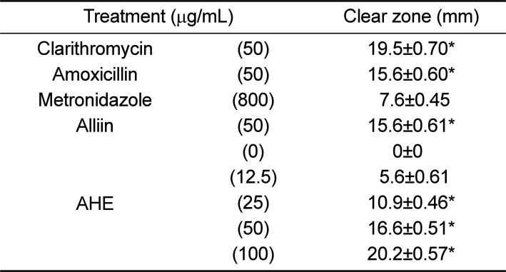
Table 1
Anti-H. pylori activities of Allium hookeri extract (AHE) using the disk agar diffusion method
![]()
Rapid urease test
Repeated intragastric inoculation (1.0×10⁹ CFU/mouse, 3 times) of H. pylori to C57BL/6 mice revealed positive reaction (red color) in Campylobacter-like organism test (CLO) test on the gastric mucosa (Figure 1). The stomachs of H. pylori-infected mice orally treated with AHEs 25, 50 and 100 mg/kg/day dose during a 4 week treatment period displayed positive reaction in 70, 50, and 10%, respectively (Table 2). Also, we calculated CLO scores as described previously [16]. As shown in Figure 2, AHEs exhibited a significant decrease of CLO scores as compared to H. pylori infected group. These mean that AHE could eliminate significantly the cases of H. pylori colonization.
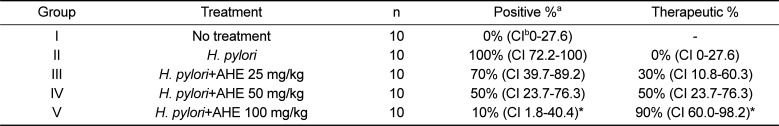
 | Figure 1Gross findings of rapid urease test with CLO test kit. (A) Negative reaction (yellow color change) with control mice. (B) Positive reaction (red color change) with H. pylori infected mice.
|
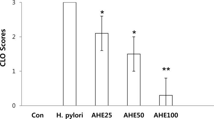 | Figure 2Score of rapid urease test with CLO test kit in H. pylori infected mice was significantly decreased by Allium hookeri extracts (AHE) (*P<0.05, **P<0.01).
|
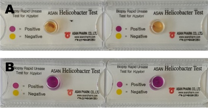
Table 2
Reactivity in CLO test on the gastric mucosa of mice infected with H. pylori followed by treatment with or without Allium hookeri extract (AHE)
![]()
Histopathological analysis
Pathological changes in the gastric mucosa were negligible in animals from non-H. pylori inoculated group I. In contrast, gastric atrophy and ulceration developed in the gastric mucosa of mice in group II (H. pylori inoculation only), indicating marked mucosal destruction. However, mice in group III, IV and V (H. pylori + AHEs 25, 50 and 100 mg/kg/day individually) revealed markedly improved the villi lesions. The histopathological lesion score in AHEs treated animals significantly lower than that in the infection control group II (H. pylori inoculation only) (Table 3).
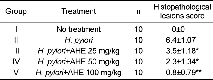
Table 3
Histopathological lesions scores of mice infected with H. pylori followed by treatment with or without Allium hookeri extract (AHE)
![]()
Go to :
Discussion
H. pylori highly colonize the human gastric mucosa and perfectly adapted to that environment. Concerning pathology, H. pylori causes gastritis and is classified as a major primary risk factor for the development of gastric or peptic ulcers, gastric adenocarcinoma and mucosa-associated lymphoid tissue lymphoma [17].
In the present study, we identified inhibition activity of the AHE against H. pylori in a mouse model. Rapid urease tests of the mice stomachs demonstrated a significant reduction in H. pylori colonization. In addition to the therapeutic effect against H. pylori infection, the AHE reduced mucosal inflammation and epithelial damages in the stomach of H. pylori-infected mice. It is likely that H. pylori eradication caused a decrease in the degree of inflammation in the stomach, although there is a possibility that AHE itself has an anti-inflammatory effect on gastric mucosa.
Allium hookeri is a wild herb cultivated in India, Myanmar, Nepal, and China. It is mainly used as food supplement and medicinal food. The beneficial effects of Allium hookeri has been extensively investigated including, anti-oxidant [18], anti-inflammatory [11], anti-microbial [19], anti-obesity [20] and antidiabetic [21]. Its beneficial effect is attributed to the sulfur compounds, phenolic compounds, phytosterols and vitamin C [22]. Allicin and ten alkyl thiosulfinates from Allium hookeri root extract were characterized by HPLC-ESIMS [23]. Non-volatile organosulfur compounds, methiin and cycloalliin were detected as major compounds and volatile compounds such as allyl methyl sulphides and dimethyl sulphides were identified from Allium hookeri [24]. Some of chemicals in Allium hookeri have showed biological activities. Ferulic acid-4-O-β-D-glucopyranoside isolated from Allium hookeri root has antioxidant activity [25]. The hot-water extract of Allium hookeri containing alliin, sinapic acid, and ferulic acid exhibited beneficial effects on bone health [26].
Although triple therapy, using two antibacterial antibiotics and a proton pump inhibitor, is effective and its short duration helps maintain patient compliance, a considerable number of patients experience undesirable side effects, such as diarrhea, epigastric pain, nausea, and bloating [27]. In contrast, Allium hookeri is relatively safe and contains a wide range of thiosulphinates such as allicin which are thought to be responsible for the antibacterial activity [212223]. These safety characteristics of AHE may therefore be appropriate for use in prevention and therapy against H. pylori infection.
In this study, AHE showed a significant inhibition effect against H. pylori infection. It could be a promising AHE treatment for patients with gastric complaints including gastric ulcers caused by H. pylori. AHE may be useful to treat patients with an H. pylori infection with high therapeutic efficacy and safety.
Go to :
 XML Download