

 PDF
PDF ePub
ePub Citation
 Print
Print


Sung Hae Park, Jun Young Lee , Hyun Woong Jang
, Hyun Woong Jang
Abstract
Purpose
To evaluate the clinical significance and usefulness of a bone scan in accessory navicular bone.
Materials and Methods
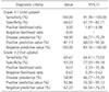
Eighty-five patients with foot pain and accessory navicular bone on radiography, who underwent bone scan from 2012 to 2015, were analyzed retrospectively. The subjects was divided into a symptomatic and asymptomatic group according to the presence of navicular bone tenderness. The grade of bone scan uptake was divided into 3 grades. Age, gender, grade of bone scan and size of the accessory navicular bone were analyzed. The symptomatic group were divided into a low (grade 0, 1) and high uptake (grade 2) group to determine the appropriate treatment. The low uptake group was treated conservatively for 3 months. The high uptake group was initially treated conservatively for 3 months and surgery was performed if pain persisted. For the clinical evaluation, the visual analogue scale, American Orthopaedic Foot and Ankle Society midfoot scale were evaluated in the first examination and last follow-up date. The patient's satisfaction grade was also evaluated at the last follow-up.
Results
The asymptomatic group mostly showed no uptake in the bone scan. On the other hand, some patients in the asymptomatic group showed an increase in uptake. In these patients, the size of accessory navicular bone was related to the grade of bone scan uptake, showing that the bone scan uptake grade can be predicted when applying different cut off values for the bone size. The symptomatic group mostly showed uptake in the bone scan and the grade of uptake had a positive correlation with the size of the accessory navicular bone (p<0.05). Age and gender were not related to the bone scan uptake. In the clinical evaluation, conservative and surgical treatment showed a good outcome.
Conclusion
The bone scan uptake grade alone cannot be used to completely predict the symptoms. On the other hand, the size of the accessory bone can increase the bone scan uptake. Therefore, the size of the accessory bone, and patient symptoms should be considered in patients with a high uptake when deciding treatment.
Figures and Tables
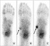 | Figure 1Grades on bone scan. Plantar images of bone scans showed grade 0 (A), grade 1 (B; arrow) and grade 2 (C; arrow).
|

Notes
References
1. Kim JR, Park CI, Moon YJ, Wang SI, Kwon KS. Concomitant calcaneo-cuboid-cuneiform osteotomies and the modified Kidner procedure for severe flatfoot associated with symptomatic accessory navicular in children and adolescents. J Orthop Surg Res. 2014; 9:131.

2. Miller TT. Painful accessory bones of the foot. Semin Musculoskelet Radiol. 2002; 6:153–161.
3. Mellado JM, Ramos A, Salvadó E, Camins A, Danús M, Saurí A. Accessory ossicles and sesamoid bones of the ankle and foot: imaging findings, clinical significance and differential diagnosis. Eur Radiol. 2003; 13:Suppl 6. L164–L177.
4. Keats TE, Anderson MW. Atlas of normal roentgen variants that may simulate disease. 9th ed. hiladelphia: Elsevier Saunders;2012.
5. Romanowski CA, Barrington NA. The accessory navicular-an important cause of medial foot pain. Clin Radiol. 1992; 46:261–264.
6. Jain S, Karunanithi S, Agarwal KK, Kumar G, Roy SG, Tripathi M. Incremental value of single photon emission tomography/computed tomography in 3-phase bone scintigraphy of an accessory navicular bone. Indian J Nucl Med. 2014; 29:191–192.
7. Veitch JM. Evaluation of the Kidner procedure in treatment of symptomatic accessory tarsal scaphoid. Clin Orthop Relat Res. 1978; (131):210–213.
8. Chiu NT, Jou IM, Lee BF, Yao WJ, Tu DG, Wu PS. Symptomatic and asymptomatic accessory navicular bones: findings of Tc-99m MDP bone scintigraphy. Clin Radiol. 2000; 55:353–355.
9. Lassmann M, Biassoni L, Monsieurs M, Franzius C, Jacobs F. EANM Dosimetry and Paediatrics Committees. The new EANM paediatric dosage card. Eur J Nucl Med Mol Imaging. 2008; 35:1748.
10. Lee J, Youn H, Choi WJ, Lee JW. Comparison of clinical outcome of excision versus osteosynthesis in type II accessory navicular. J Korean Foot Ankle Soc. 2011; 15:72–78.
11. Chisin R, Peyser A, Milgrom C. Bone scintigraphy in the assessment of the hallucal sesamoids. Foot Ankle Int. 1995; 16:291–294.
12. Geist ES. The accessory scaphoid bone. J Bone Joint Surg. 1925; 7:570–574.
13. Mygind HB. The accessory tarsal scaphoid; clinical features and treatment. Acta Orthop Scand. 1953; 23:142–151.
 XML Download