

 PDF
PDF ePub
ePub Citation
 Print
Print


INTRODUCTION
Ectropion is defined as an outward rotation of the eyelid margin and may be divided into the following categories: congenital, involutional, paralytic, and cicatricial1. Paralytic ectropion is commonly seen in patients with a trauma of the facial nerve, Bell palsy, central nervous system tumors, and leprosy. Leprosy is a chronic granulomatous infection caused by Mycobacterium leprae, which affects primarily skin and nerves2. In patients with leprosy, paralysis of the facial nerve is associated with decreased muscle tone and lateral canthal tendon laxity, disturbing the orbicularis oculi's closing the eyelids, and finally resulting in epiphora (overflow of tears), the lower eyelid ectropion, entropion (an inversion of the eyelid) and lagophthalmos (inability to close the eyes fully)3. Furthermore, these changes can cause exposure keratitis and corneal ulceration, leading to even blindness123. Treatment of paralytic ectropion is usually surgery. One of surgical techniques which involves tightening the lateral canthal tendon is called lateral canthoplasty or lateral tarsal strip (LTS) procedure, which is commonly performed by ophthalmologists but rarely by dermatologic surgeons13. According to Lewallen et al.4, about 34% of patients with leprosy developed ocular lesions during course of their disease. Although the dermatologists are primary physicians to diagnose and treat patients with leprosy, not many dermatologists are aware of such complications or know how to treat them.
Herein we retrospectively analyze the efficacy of the LTS for correction of paralytic ectropion in patients with leprosy and review the leprosy related ocular complication.
MATERIALS AND METHODS
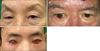
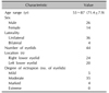
A total of 40 Korean patients with paralytic ectropion who had visited Korean Hansen Welfare Association Hospital were treated with the LTS between January 2010 and December 2015. All of them were cured leprosy patients, bacteriologically negative with morphological indices of 0%, and did not respond to conservative treatments such as lubricant eye drops and ointments. Patients were classified into 4 ectropion severity categories (mild, moderate, marked, and extreme) using a grading system designed by Moe and Linder (Table 1, Fig. 1)5.
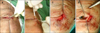
The surgical technique involves a lateral canthotomy extending 1 cm to the lateral orbital rim using knife or scissors (Fig. 2A). A lateral cantholysis is performed by incising the inferior crus of the lateral canthal tendon. The eyelid is split into anterior and posterior lamellae. A tarsal strip is fashioned by trimming off the mucocutaneous junction superiorly (Fig. 2B). Once having fashioned the tarsal strip, it was shortened by the amount necessary. Then the tarsal strip is grasped with a forcep and the conjunctival surface is scraped using a No. 15 blade to clean the epithelium from the tarsal strip. The tarsal strip is sutured tightly to the periosteum on the inner aspect of the lateral orbital rim superior to the lateral canthal tendon insertion with a 5-0 nylon (Fig. 2C). The lateral canthotomy is closed by layering suture of conjunctiva and the anterior lamella after excision of redundant skin (Fig. 2D).
Local complications including postoperative hemorrhage, wound infection, and wound dehiscence, 4-point patients' global assessment scale on improvement of symptoms such as epiphora, redness, discharge, sensation of foreign body, ability to close eyelids (patient global assessment of disease activity; PtGA, 0: poor, 1: fair, 2: good, 3: excellent), and recurrence rate were assessed after one year of follow-up. The patients were asked if the symptoms listed above improved after the surgery and if the symptoms showed 0%~25% improvement, PtGA of 0 (poor) was given, 26%~50%, 1 (fair), 51%~75%, 2 (good), 76%~100%, 3 (excellent). The study protocal was approved by the institutional review boards of the Veterans Health Service Medical Center (IRB no. 2016-09-008).
RESULTS
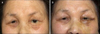
The characteristics of the patients are summarized in Table 2. The patients, 26 male and 14 female, were between the ages of 53 and 87 years (mean 71.4±7.9 years) and the total number of eyelid operation was 44 because of bilateral lesions in 4 patients. Eyelids of most patients showed moderate degree of ectropion (n=35); 5 eyelids showed mild degree, 4 eyelids showed marked degree, and none had extreme degree of ectropion. Recurrence was observed in 5 eyelids (5/44, 11.4%) during 1 year follow-up period. Of 5, 2 eyelids were marked degree of ectropion (2/3, 66.7%) and 3 had moderate degree of ectropion (3/32, 9.4%). The patients with mild degree of ectropion showed no recurrence. Most patients were satisfied with the results (Fig. 3, 4) and mean PtGA scale was 2.6 at the end of follow-up period. There were no serious postoperative complications except mild size discrepancy of about 1~2 mm in both eyes in 2 patients.
DISCUSSION
Ocular damage in leprosy patients is caused by nerve damage or mycobacterial infiltration. Ocular complications of leprosy occur from infiltration of M. leprae in fifth and seventh cranial nerve, eyes, and surrounding tissues, causing secondary changes in eyelids, cornea, uvea, and lens. The pathogenesis of ocular damage in leprosy can be simplified by differentiating the process into the tuberculoid and lepromatous categories3. In tuberculoid state, inflammatory patch over the facial nerve which innervates the orbicularis oculi muscles can result in eyelid dysfunction. In lepromatous state, direct invasion of M. leprae in the nerve and ocular structures occurs, putting the leprosy patients at highest risk for all of the ocular complications3. As in other parts of the body, the cooler area in the anterior part of the eye and the surrounding tissues are more susceptible to the invasion of M. leprae, which result in leprosy-related ocular complications including madarosis, impaired lid closure, iris atrophy, and keratitis3. Small branches of the trigeminal nerve which innervate the cool, avascular corneal structures become targets of the leprosy bacilli and result in decreased ocular sensation. And as cornea becomes less sensitive, it is more prone to injuries and corneal opacity and cataract are more likely to occur. Zygomatic branch of facial nerve, responsible for innervating orbicularis oculi muscle, crosses the malar eminence of the zygomatic bone in a thin layer of tissue cooler than the surrounding areas, which makes attack of the leprosy bacilli against this branch more vulnerable than others due to the cooler temperature23. Involvement of this branch results in paralysis of orbicularis oculi muscle, responsible for closing of eyelids, and lead to lagophthalmos, ectropion, and entropion. Paralysis can be wholly or partially prevented by antibacterial chemotherapy; however, once paralysis is established, recovery is usually minimal to none at all. Between 3 and 19.8% of leprosy patients suffer from facial nerve palsy6.
Paralytic ectropion can easily be diagnosed by simple physical examination; however, the functional and cosmetic problems are difficult to manage, and may even lead to blindness if left untreated. The main goal of therapy is to keep the cornea moist and the simplest way in doing so is using eye lubricants such as eyedrops and ointments. Other treatment options include physical therapy which is teaching patients to forcefully close their eyes several times a day or taping the eyes closed at night has been tried3. Surgery can be considered in cases of exposure keratitis where conservative treatments are no longer helpful, and it can also be considered for cosmetic purposes. Conservative treatments can be used to prevent severe complications, however, eyelid surgery is the only treatment which consistently gives satisfactory results36. The most common surgical technique for repair of ectropion is LTS, which is a quick and easy procedure. Kim et al.7 conducted a similar study and reported that about 90% of leprosy patients who underwent LTS were satisfied with the results. And in our study, most patients were satisfied with the results and mean PtGA scale was 2.6/3 at the end of follow-up period, which was similar to those of ophthalmologists. However, in very lax eyelids, surgery confined to the lateral canthal tendon has not been able to correct all the laxity. Therefore, preoperative evaluation of lower eyelid ectropion is critical in every patient with leprosy before surgical planning. A grading system designed by Moe and Linder5 is helpful, and we, on the basis of this system, classified ectropion severity into 4 categories: mild, moderate, marked, and extreme. Our patients were mostly in moderate degree and no one was in extreme degree. Whereas the LTS procedure is suited for mild and moderate degree of ectropion, patients with marked and extreme degree need additional or more radical methods such as wedge resection in the former and temporalis muscle transfer in the latter8. Recurrence was seen in 5 eyelids and the recurrence rate was 11.4% (5/44). While the patients with mild and moderate degree of ectropion showed recurrence of 0% (0/5) and 9.4% (3/32) respectively, 66.7% (2/3) of the patients with marked degree of ectropion recurred during follow-up period. Thus, the LTS is much more suitable for mild to moderate degree of paralytic ectropion, and other or additional procedure is necessary for the patients with marked degree of ectropion. Since the patients showing recurrence in moderate degree of ectropion were in late eighth and ninth decades, senile change may be related to recurrence.
The details of surgical technique of the LTS procedure is slightly different in several articles9101112131415, but the common final goal is the correction of eyelid malposition by horizontal tightening and canthal elevation with formation of a new lateral canthus. If several important points such as a firm fixation to the periosteum, removal of conjunctiva inside the tarsal strip, and a slight overcorrection in both tightening and canthal elevation are emphasized12, the goal can be accomplished easily. But canthal angles in both eyes should be similar.
The complications were few and there were no serious complications such as infection, amputation of tarsal strip, point tenderness of anchoring site, or suture granulomas13. Mild size discrepancy of both eyes was observed in 2 patients probably due to overcorrection one year after surgery, but most patients were satisfied (mean PtGA scale was 2.6/3). The study limitations include its retrospective nature, a single institution and a single dermatologic surgeon, and relatively brief follow-up period. Thus, clinical information regarding the subtypes of leprosy, duration of the disease, and treatment history, which might have affected the outcome of the surgery, could not be included. And the single-surgeon study design could also be a limitation as the results may not be representative of a large cohort of surgeons' experience, and the outcomes were evaluated based on patients' satisfactory rates. Leprosy patients have limited access to general health services as they tend to live in leprosy villages or settlements. And even in leprosy treatment facilities, full-time ophthalmologists are not always available, making it difficult for patients to receive ophthalmologic evaluations. A simple, easy grading system along with safe and effective LTS showed satisfactory results in repair of mild and moderate degrees of paralytic ectropion. We hope the results of our study will encourage the dermatologists who play pivotal roles in the management of leprosy to participate more in screening and treatment of ocular complications in leprosy patients.
In conclusion, the LTS along with simple, easy grading system is a simple, safe, and effective treatment method for the dermatologic surgeons to correct paralytic ecectropion of mild and moderate degree in patients with leprosy.

 XML Download