

 PDF
PDF ePub
ePub Citation
 Print
Print


Dear Editor:
Vitiligo is an acquired depigmenting disorder caused by the loss of melanocytes. Vitiligo universalis is complete or nearly complete depigmentation of the entire body surface1. We herein report a case of repigmentation in a vitiligo universalis patient.
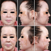
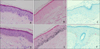
A 50-year-old woman, diagnosed with vitiligo universalis 20 years ago, presented with multiple, hyperpigmented macules and patches on the face for 5 months (Fig. 1). She had not received any treatment for vitiligo. She had sigmoid colon cancer and had 10 cycles of FOLFOX chemotherapy consisting of oxaliplatin, fluorouracil, and leucovorin for 4 months. The pigmented lesions on the face were said to have developed after starting chemotherapy. We initially considered a diagnosis of melasma, lentigines, or postinflammatory hyperpigmentation. Histopathological findings of the hypopigmented lesion showed loss of melanocytes and melanin pigment. The hyperpigmented macule revealed increase of basal melanin pigment and melanocytes (Fig. 2). These histopathological results suggested the diagnosis of repigmentation of vitiligo universalis. A depigmentation cream (Melanon cream, Dong-A Pharm., Seoul, Korea) was given.
Most studies report that melanocytes are completely absent in vitiligo skin, suggesting that only hair follicles can act as a reservoir. However, recent reports provide evidence that melanocytes can recover their functionality in vivo and in vitro under appropriate stimulus2,3,4.
Repigmentation in vitiligo may also occur spontaneously and may be therapy-induced. Spontaneous repigmentation is unpredictable and occurs in less than 15%∼25% of patients1.
Ultraviolet radiation can also stimulate melanocyte activity. Sun-exposed areas are more favorable for repigmentation with about twofold higher density of melanocytes than in non-exposed areas3. However, our patient was a housekeeper and had very little sun exposure.
The onset of repigmentation after chemotherapy in our patient questioned the effect of chemotherapy on repigmentation. There has been a report of repigmentation in a vitiligo universalis patient treated for pemphigus vulgaris with dexamethasone cyclophosphamide pulse therapy3. This report suggested that systemic immunossupressive drugs, including corticosteroids, which are used to treat vitiligo, could be responsible for initiating melanocyte activity3.
Among the FOLFOX regimen, fluorouracil can cause variable patterns of hyperpigmentation, including diffuse, mottled, reticulate pigmentation, and serpiginous supravenous hyperpigmentation5. It is suggested that fluorouracil could increase pigmentation by means of melanocyte-stimulating hormone or by direct stimulation of melanocytes themselves5.
In our patient, one month after stopping chemotherapy, the initial pigmented lesions lightened, which indicate that chemotherapy could have affected repigmentation. However, because new pigmented lesions appeared despite stopping chemotherapy, spontaneous repigmentation cannot be completely ruled out.
It is necessary to follow up several months after the withdrawal of chemotherapy due to the residual and cumulative effect of the drug. If new pigmented lesions appear even 5 to 6 months after stopping chemotherapy, spontaneous repigmentation is more likely to be the cause of repigmentation than chemotherapy. Unfortunately, our patient was lost for further follow-up.
When pigmented lesions appear in vitiligo universalis patients, it is easy to consider pigmented skin disorders such as melasma2. Sudden repigmentation of vitiligo universalis is a rare event that must be evaluated carefully to avoid misdiagnosis.
 XML Download