

 PDF
PDF ePub
ePub Citation
 Print
Print


Dear Editor:
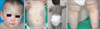
We report the case of an 18-month-old Korean female paitent. The patient presented to our clinic with congenital erythematous lesions on her face with large bluish-gray patches on the trunk and extremities. She was born full-term through a cesarean section, weighed 4,400 g, and her mother experienced no pathological events during pregnancy and delivery. The familial history was unremarkable for lesions. The general physical examination revealed, bilateral nevus of Ota with melanosis bulbi on her face and generalized port-wine stains (nevus flammeus) on her whole body. In addition, extensive bluish-gray hyperpigmented patches (Mongolian spots) were located on the trunk and extremities (Fig. 1). On the developmental evaluation, she presented mild developmental delay (The patients could stand alone but could not walk alone perfectly). The right thigh circumference was smaller than the left thigh circumference by 2 cm, but this sign was within normal variation range. Magnetic resonance imaging of the brain did not reveal abnormal findings.
Phacomatosis pigmentovascularis (PPV) is a rare disorder characterized by the combination of vascular malformation and pigmentary abnormalities. In 1947, Ota et al.1 first described PPV as a disorder characterized by the combination of melanocytosis and capillary malformation. Since then approximately 250 cases have been reported. Hasegawa and Yasuhara2 classified PPV into four types according to the combination of pigmentary skin lesions and nevus flammeus. First described by Trrelo et al. in 2003, type V is characterized by cutis marmorata telangiectatica congenita associated with Mongolian nevi3. Each types is subdivided according to oculocutaneous manifestation only (subtype a), or extracutaneous manifestations (subtype b) (Table 1). The major extracutaneous manifestations are neurological and skeletal symptoms, and the most common syndromes associated with PPV are Sturge-Weber syndrome and Klippel-Trenaunay syndrome. The diagnosis of PPV is primarily clinical. Type IIb (45%) is the most common type of PPV, followed by IIa (30%)4. The pathogenesis of PPV remains unclear; it has been proposed as an abnormality in the development of melanocytic nevus cells and vasomotor neural cells. Didymosis, a phenomenon known as “twin spot” is an accepted etiology of PPV35. Our patient presented with nevus flammeus and aberrant Mongolian spots. Therefore, we classified her condition as a type IIa PPV, and the patient is currently being followed for development of any systemic symptoms.
PPV without extracutaneous involvement has a benign course and does not require treatment. For cosmetic purposes, Q-switched pulsed dye laser and Q-switched Nd-Yag laser can be helpful for the vascular component and nevus component, respectively. To our knowledge, PPV is rarely reported in Korean patient. Herein we report the case of PPV type IIa diagnosed in an 18-month-old female patient.

 XML Download