

 PDF
PDF ePub
ePub Citation
 Print
Print


Abstract
Purpose
We report a case of late-onset capsular block syndrome, which resulted in a misdiagnosis of intraocular lens (IOL) opacity.
Case summary
A 59-year-old man visited our clinic with reduced visual acuity in the right eye from 1 year prior. He had undergone uncomplicated bilateral cataract surgery by phacoemulsification with IOL implants at another hospital 10 years before. There was no specific history with the exception of hypertension. After being diagnosed in the ophthalmology clinic with IOL degeneration and opacity in the right eye, he was referred to our hospital for IOL replacement. Upon examination, his right uncorrected visual acuity was 0.06 and intraocular pressure was 22 mmHg. The refractive error could not be checked due to IOL opacity. Slit-lamp microscopy revealed a cloudy, milky IOL. Anterior-segment optical coherence tomography of the right eye showed retention of a highly reflective material in the lens capsule behind the IOL. Posterior capsule enlargement of the right eye was confirmed on ultrasound biomicroscopy. After neodymium-doped yttrium aluminium garnet (Nd:YAG) laser capsulotomy was performed, the homogeneous space disappeared and the eye recovered normal visual acuity.
Conclusions
Capsular block syndrome is a rare complication that can occur shortly (1 day to 2 days) after cataract surgery. Late-onset capsular block syndrome, which occurs 10 years after surgery differs from typical clinical manifestations. Thus, capsular block syndrome is an important consideration upon the presentation of opacification due to IOL degeneration.
Figures and Tables
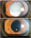 | Figure 1Slit lamp photography of the right eye. (A) The anterior capsular incision was fibrous and adhered to the optic part of intraocular lens (IOL), cloudy milky material was observed in the intraocular lens space behind the IOL. And the posterior capsule was swollen. (B) After the neodymium-doped yttrium aluminium garnet (Nd:YAG) capsulotomy, milky white material was not observed behind the intraocular lens space and the uncorrected visual acuity was 1.0.
|
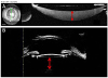 | Figure 2Anterior optical coherence tomography and ultrasound biomicroscopy. (A) Anterior segment optical coherence tomography of the right eye showed retention of highly reflective material in the lense capsule behind the intraocular lens. Arrow showed Retention of highly reflective material in the capular bag. (B) Posterior capsule enlargement of right eye was confirmed on ultrasound biomicroscopy. Arrow showed capsular bag distension. IR = infrared; ART = automatic real-time tracking; OCT = optical coherence tomography; HR = high resolution.
|
Notes
References
1. Miyake K, Ota I, Ichihashi S, et al. New classification of capsular block syndrome. J Cataract Refract Surg. 1998; 24:1230–1234.

2. Heo JY, Ahn MD, Joo CK. Two cases of late postoperative capsular block syndrome. Korean J Ophthalmol. 1999; 13:105–109.
3. Rana M, Jiang L, Ilango B, Yang YC. Late-onset capsular block syndrome: unusually delayed presentation. Case Rep Ophthalmol. 2013; 4:299–302.
4. Kim HK, Shin JP. Capsular block syndrome after cataract surgery: clinical analysis and classification. J Cataract Refract Surg. 2008; 34:357–363.
5. Davison JA. Capsular bag distension after endophacoemulsification and posterior chamber intraocular lens implantation. J Cataract Refract Surg. 1990; 16:99–108.
6. Holtz SJ. Postoperative capsular bag distension. J Cataract Refract Surg. 1992; 18:310–317.
7. Choi BJ, Jin YB, Hur J. Postoperative capsular bag distension in cataract surgery. J Korean Ophthalmol Soc. 1998; 39:2031–2037.
8. Lee SJ, Kim HY, Oum BS, Lee JE. Early capsular block syndrome after phacoemulsification with posterior chamber IOL insertion combined with vitrectomy. J Korean Ophthalmol Soc. 2013; 54:716–722.
9. Basti S, Nayak H, Mathur U. Capsular bag distension after optic captureof a sulcus-fixated intraocular lens. J Cataract Refract Surg. 1999; 25:293–295.
10. Geyer O, Goldstein M, Rothkoff L, Lazar M. Capsular bag distension associated with sulcus implantation of intraocular lenses. J Cataract Refract Surg. 1998; 24:1538–1540.
11. Sugiura T, Miyauchi S, Eguchi S, et al. Analysis of liquid accumulated in the distended capsular bag in early postoperative capsular block syndrome. J Cataract Refract Surg. 2000; 26:420–425.
12. Sorenson AL, Holladay JT, Kim T, et al. Ultrasonographic measurement of induced myopia associated with capsular bag distention syndrome. Ophthalmology. 2000; 107:902–908.
13. Eifrig DE. Capsulorhexis-related lacteocrumenasia. J Cataract Refract Surg. 1997; 23:450–454.
14. Qu J, Bao Y, Li M, et al. Surgical management of late capsular block syndrome. J Cataract Refract Surg. 2010; 36:1687–1691.
15. Neuhann IM, Werner L, Izak AM, et al. Late postoperative opacification of a hydrophilic acrylic (hydrogel) intraocular lens: a clinicopathological analysis of 106 explants. Ophthalmology. 2004; 111:2094–2101.
16. Bompastor-Ramos P, Póvoa J, Lobo C, et al. Late postoperative opacification of a hydrophilic-hydrophobic acrylic intraocular lens. J Cataract Refract Surg. 2016; 42:1324–1331.
17. Ramos JL, Li Y, Huang D. Clinical and research applications of anterior segment optical coherence tomography - a review. Clin Exp Ophthalmol. 2009; 37:81–89.
18. Kucukevcilioglu M, Hurmeric V, Erdurman FC, Ceylan OM. Imaging late capsular block syndrome: ultrasound biomicroscopy versus Scheimpflug camera. J Cataract Refract Surg. 2011; 37:2071–2074.
 XML Download