

 PDF
PDF ePub
ePub Citation
 Print
Print


Abstract
Purpose
To assess the risk of development of secondary glaucoma after congenital cataract surgery using a long-term fol-low-up study.
Methods
In total, 148 eyes of 91 patients who underwent congenital cataract surgery at our hospital or other hospitals were included in a retrospective chart review. A diagnosis of secondary glaucoma was made if the intraocular pressure (IOP) exceeded 21 mmHg and the corneal diameter, axial length, or the cup-to-disc ratio increased, or surgery was performed to control the IOP. To analyze the clinical features and risk factors of secondary glaucoma, we evaluated the mean age at cataract surgery, binoc-ularity, presence of a nuclear cataract, methods of cataract surgery, presence of an intraocular lens (IOL), duration of diagnosis of secondary glaucoma after cataract surgery, duration of follow-up, recent best-corrected visual acuity, and refractive errors.
Results
Thirty-five eyes (23.6%) were diagnosed with secondary glaucoma as a complication of congenital cataract surgery. Of these, 11 eyes (31.4%) were treated with glaucoma surgery a mean of 3.4 times. The mean duration from congenital cataract surgery to diagnosis of glaucoma was 112.2 ± 113.1 months. Patients with aphakia had a higher risk of developing secondary glaucoma compared with patients undergoing primary IOL implantation (p = 0.001). Younger age (<3 months at surgery), a nuclear cataract, and aphakia were risk factors for the development of secondary glaucoma (p = 0.03, p = 0.006, and p < 0.001, respectively), and the risk of developing secondary glaucoma increased with secondary IOL implantation (p = 0.052).
Conclusions
Secondary glaucoma after congenital cataract surgery was more common in patients with secondary IOL implantation, aphakia, a younger age (<3 months), and a nuclear cataract. Patients who underwent congenital cataract surgery had an increased risk for developing secondary glaucoma. Long-term monitoring of the IOP and optic nerve is therefore required for these patients.
References
1. Asrani S, Freedman S, Hasselblad V, et al. Does primary abdominal lens implantation prevent “aphakic” glaucoma in children? J AAPOS. 2000; 4:33–9.
2. Kang KD, Yim HB, Biglan AW. Comparison of delayed-onset glaucoma and early-onset glaucoma after infantile cataract surgery. Korean J Ophthalmol. 2006; 20:41–6.

3. Mills MD, Robb RM. Glaucoma following childhood cataract surgery. J Pediatr Ophthalmol Strabismus. 1994; 31:355–60. abdominal 361.
4. Lundvall A, Zetterström C. Complications after early surgery for congenital cataracts. Acta Ophthalmol Scand. 1999; 77:677–80.
5. Chak M, Rahi JS; British Congenital Cataract Interest Group. Incidence of and factors associated with glaucoma after surgery for congenital cataract: findings from the British Congenital Cataract Study. Ophthalmology. 2008; 115:1013–8.e2.
6. Kirwan C, Lanigan B, O'Keefe M. Glaucoma in aphakic and abdominal eyes following surgery for congenital cataract in the first year of life. Acta Ophthalmol. 2010; 88:53–9.
7. Trivedi RH, Wilson ME, Golub RL. Incidence and risk factors for glaucoma after pediatric cataract surgery with and without abdominal lens implantation. J AAPOS. 2006; 10:117–23.
8. Beck AD, Freedman SF, Lynn MJ, et al. Glaucoma-related adverse events in the Infant Aphakia Treatment Study: 1-year results. Arch Ophthalmol. 2012; 130:300–5.
9. Freedman SF, Lynn MJ, Beck AD, et al. Glaucoma-Related Adverse Events in the First 5 Years After Unilateral Cataract Removal in the Infant Aphakia Treatment Study. JAMA Ophthalmol. 2015; 133:907–14.
10. El Shakankiri NM, Lotfy Bayoumi NH. The timing of surgery for congenital cataracts: delayed surgery for best surgical outcomes. J AAPOS. 2016; 20:192–3.
11. Kuhli-Hattenbach C, Lüchtenberg M, Kohnen T, Hattenbach LO. Risk factors for complications after congenital cataract surgery without intraocular lens implantation in the first 18 months of life. Am J Ophthalmol. 2008; 146:1–7.
12. Cape CJ, Zaidman GW, Beck AD, Kaufman AH. Phenotypic variation in ophthalmic manifestations of MIDAS syndrome (microphthalmia, dermal aplasia, and sclerocornea). Arch Ophthalmol. 2004; 122:1070–4.
13. Rabiah PK. Frequency and predictors of glaucoma after pediatric cataract surgery. Am J Ophthalmol. 2004; 137:30–7.
14. Swamy BN, Billson F, Martin F, et al. Secondary glaucoma after paediatric cataract surgery. Br J Ophthalmol. 2007; 91:1627–30.
15. Lambert SR, Purohit A, Superak HM, et al. abdominal risk of abdominal after congenital cataract surgery. Am J Ophthalmol. 2013; 156:355–61.e2.
16. Sukhija J, Kaur S, Ram J. Outcome of primary intraocular lens abdominalation in infants: Complications and rates of additional surgery. J Cataract Refract Surg. 2016; 42:1060–5.
17. Mataftsi A, Haidich AB, Kokkali S, et al. Postoperative glaucoma following infantile cataract surgery: an individual patient data meta-analysis. JAMA Ophthalmol. 2014; 132:1059–67.
18. Magnusson G, Abrahamsson M, Sjöstrand J. Glaucoma following congenital cataract surgery: an 18-year longitudinal follow-up. Acta Ophthalmol Scand. 2000; 78:65–70.
19. Wong IB, Sukthankar VD, Cortina-Borja M, Nischal KK. Incidence of early-onset glaucoma after infant cataract extraction with and without intraocular lens implantation. Br J Ophthalmol. 2009; 93:1200–3.
20. Chen W, Long E, Chen J, et al. Timing and approaches in abdominal cataract surgery: a randomised controlled trial. The Lancet. 2016; 388:S52.
21. Kumar M, Arora R, Sanga L, Sota LD. Scleral-fixated intraocular lens implantation in unilateral aphakic children. Ophthalmology. 1999; 106:2184–9.
22. Jacobi PC, Dietlein TS, Jacobi FK. Scleral fixation of secondary foldable multifocal intraocular lens implants in children and young adults. Ophthalmology. 2002; 109:2315–24.
23. Pakravan M, Homayoon N, Shahin Y, Ali Reza BR. Trabeculectomy with mitomycin C versus Ahmed glaucoma implant with mitomycin C for treatment of pediatric aphakic glaucoma. J Glaucoma. 2007; 16:631–6.
Figure 1.
Kaplan-Meier curves showing cumulative probability of an eye's developing glaucoma or glaucoma suspect after congenital cataract surgery over times. The probability of development glaucoma or glaucoma suspect was estimated to be 38.2 ± 0.1% by 20 years after congenital cataract surgery.
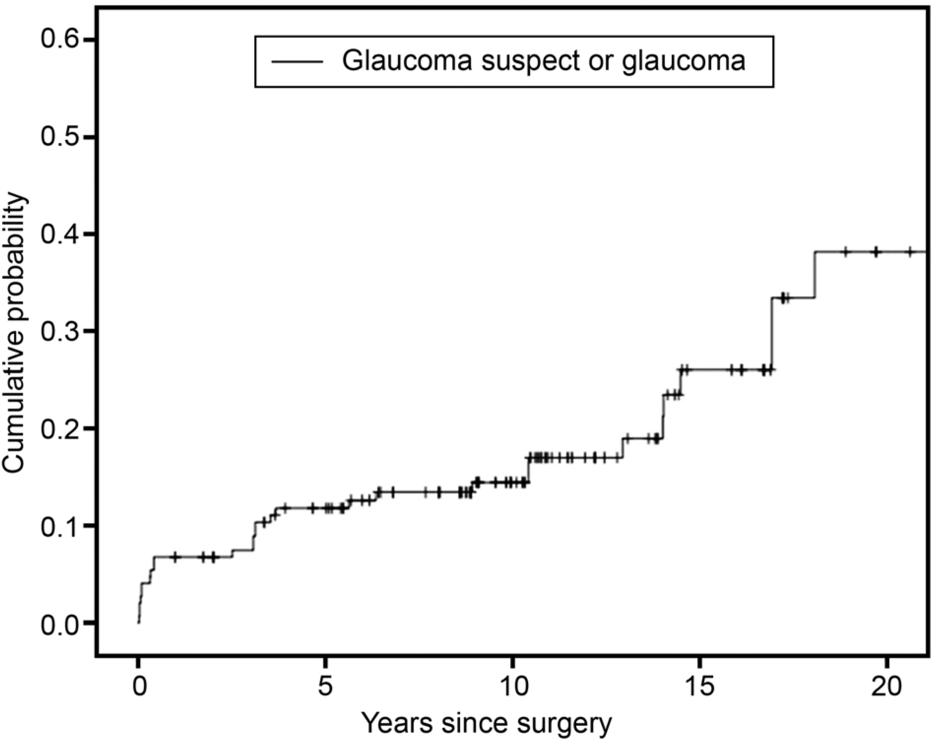
Figure 2.
Kaplan-Meier curves showing cumulative probability of an eye's developing glaucoma or glaucoma suspect after congenital cataract surgery between each group; primary intraocular lens (IOL) implantation, secondary IOL implantation and aphakia. There were statistical differences of developing glaucoma by logrank test (p < 0.001).
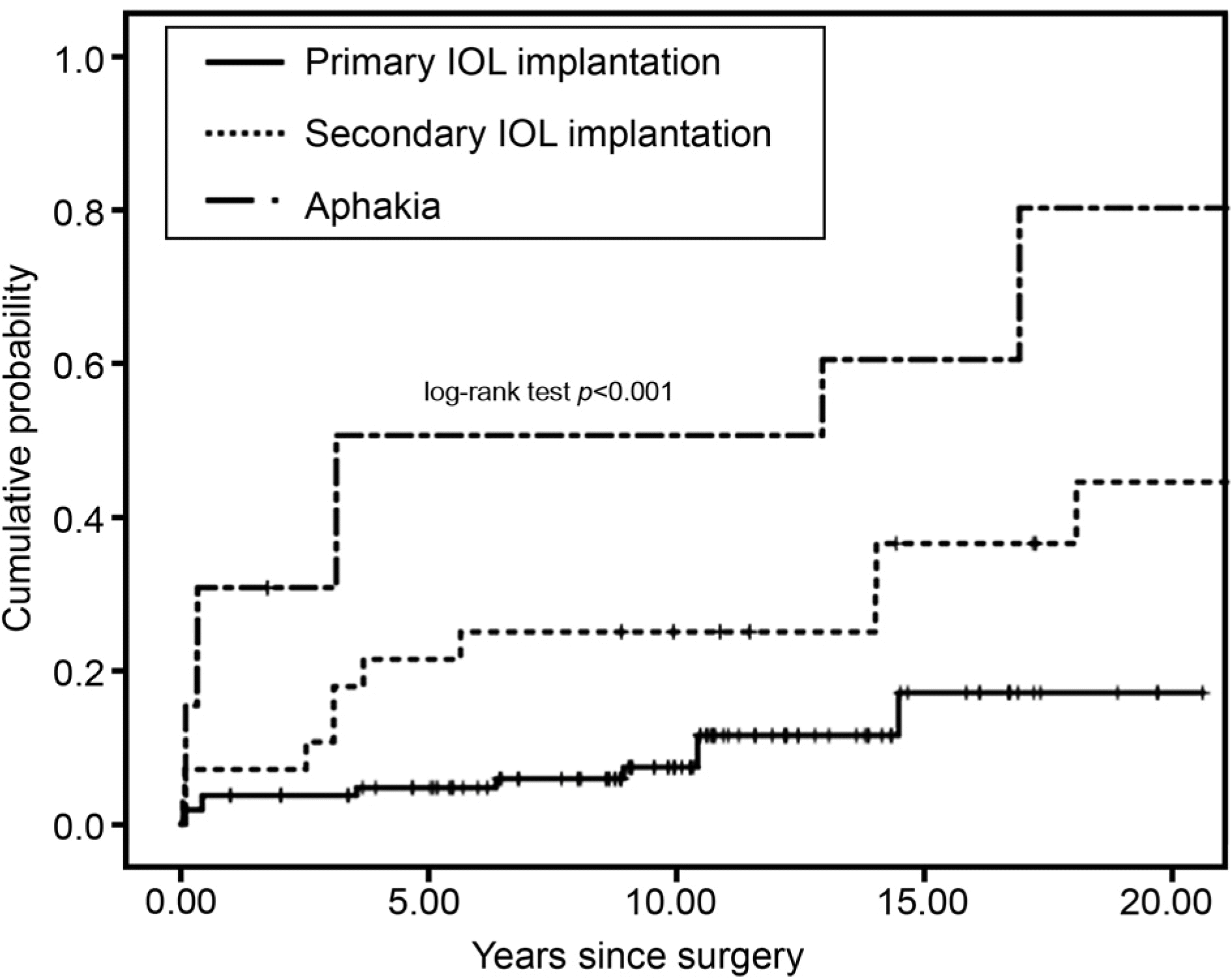
Figure 3.
Angle photo of a case with secondary glaucoma associated with congenital cataract at the time of glaucoma surgery. Ciliary body band was not visible and scleral spur was partially observed.
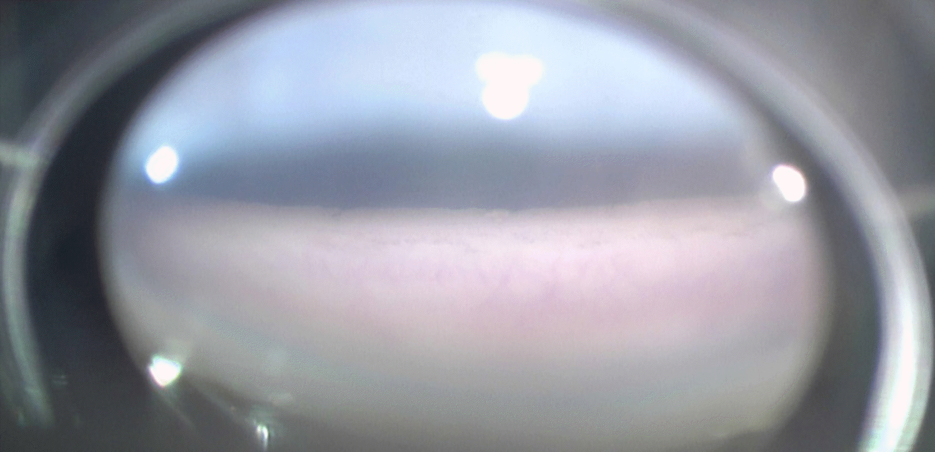
Table 1.
Demographics of 148 eyes with congenital cataract
Table 2.
Type of cataract surgical procedures
Table 3.
Diagnosis with glaucoma and glaucoma suspect
Table 4.
Univariate cox regression analysis of potential predictors to secondary glaucoma after congenital cataract surgery
| Parameter | Hazard ratio (95% CI) | p-value |
|---|---|---|
| Sex | – | 0.879 |
| Cataract surgery before 3 months of birth | 2.59 (1.05–6.40) | 0.040 |
| Nuclear cataract | 3.80 (1.40–10.28) | 0.008 |
| Bilaterality | – | 0.340 |
| Anterior vitrectomy | – | 0.350 |
| Secondary IOL implantation* | 3.16 (1.33–7.50) | 0.009 |
| Aphakia* | 7.37 (2.97–18.30) | <0.001 |
Table 5.
Univariate cox regression analysis of potential predictors to secondary glaucoma in variable surgical methods
| Parameter | Hazard ratio (95% CI) | p-value |
|---|---|---|
| Primary IOL implantation in the bag* | ||
| Primary IOL implantation in the sulcus | 4.36 (0.39–48.24) | 0.230 |
| Primary IOL implantation c scleral fixation | 101.29 (13.16–779.92) | <0.001 |
| Primary IOL implantation c piggybacking | 5.87 (1.13–30.58) | 0.035 |
| Secondary IOL implantation in the bag | 11.52 (2.18–60.86) | 0.004 |
| Secondary IOL implantation in the sulcus | 11.19 (2.0–62.5) | 0.006 |
| Secondary IOL implantation c scleral fixation | 7.93 (1.48–42.40) | 0.017 |
| Aphakia | 23.32 (5.02–108.29) | <0.001 |
Table 6.
Multivariate cox analysis of potential predictors to secondary glaucoma after congenital cataract surgery
| Parameter | Hazard ratio (95% CI) | p-value |
|---|---|---|
| Cataract surgery before 3 months of birth | 6.31 (1.18–33.70) | 0.030 |
| Nuclear cataract | 7.40 (1.78–30.70) | 0.006 |
| Primary IOL implantation* | ||
| Secondary IOL implantation | 4.82 (0.99–23.54) | 0.052 |
| Aphakia | 29.32 (4.22–203.70) | <0.001 |
 XML Download