This article has been
cited by other articles in ScienceCentral.
Dear Editor:
A 40-year-old man presented with a nodule on the extensor surface of his left forearm. He couldn't remember when it formed. Initially, it showed no symptoms but with time, became painful. Physical examination revealed a slightly bluish to skin colored, hard mobile nodule on the left forearm measuring 1×1 cm in size (
Fig. 1A). The initial clinical suspicion was epidermal cyst, trichilemmal cyst, or lipoma; thus, ultrasonography (US) was done. US results indicated a well-defined oval isoechoic mass in the subcutaneous fat layer, with moderate hyperemia (
Fig. 1B). A complicated epidermoid cyst, or other hypervascular mass, such as vascularized leiomyoma or (least likely) a sarcoma (e.g., malignant fibrous histiocytoma) was suspected. Excisional biopsy was performed. Microscopic examination showed a deep, well-defined tumor with basophilic lobules, encapsulated by thick connective tissue in the dermis and subcutaneous layer. A trabecular arrangement of basophilic cells was present (
Fig. 1C). Two types of cells (small, dark, basaloid cells with hyperchromatic nuclei, and cells with large, pale, ovoid nuclei) were seen (
Fig. 1D). The diagnosis was confirmed as eccrine spiradenoma (ES). The tumor was completely excised.
ES is a rare benign soft tissue tumor first described by Kersting and Helwig (1956). It may occur at any age, but typically affects young adults aged 15~35 years, with no sexual predilection
1. It presents as a solitary mass localized in the skin and subcutaneous tissue, smaller than 1 cm in size. Paroxysmal pain and tenderness may often present
2. It tends to arise on the upper part of the body; head, neck, and trunk, especially ventral portion
1. Rarely, it may be located on the upper and lower extremities, especially dorsal portion
3. The summarization of the previously reported cases on extremities was described in
Table 1. ES lesions are mostly solitary, but rarely multiple, linear, blaschkoid, or grouped. The pathogenesis is thought to be related to differentiation, mainly of the secretory portion of the secretory coil of eccrine sweat glands
24. The presence of myoepithelial cells and phosphorylase, demonstrate the eccrine origin of the tumor. However, several evidences suggest its origin is apocrine rather than eccrine
4. It is sometimes associated with trichoepithelioma and cylindroma, tumors of apocrine or folliculosebaceous-apocrine origin. US study revealed a well-demarcated mass with lobulated contours, which didn't have any relation with the epidermis and didn't extend into the neighboring muscular tissues. It was localized in the superficial part of the subcutaneous tissue
35. When the mass shows well-defined hypoechogenicity, hypervascularity, no cystic portion, no hypoechoic scar, and when it is deeply located, an ES can be highly considered
5. The essential histologic features include sharply demarcated nodules of basaloid cells in the dermis or in subcutaneous tissue (blue balls)
5. Basaloid cells are composed of two types which might not be so apparent: one is paler, with more cytoplasm than in the darker cells
1. Malignant change is rare
2. However, in cases with incomplete surgical removal, a high risk of local recurrence has been reported
25. Early and complete surgical excision is the diagnostic tool and curative treatment option for ES
2.
Figures and Tables
Fig. 1
(A) A solitary, mild bluish to skin-colored, hard mobile nodule on the extensor surface of the left forearm measuring 1×1 cm (black arrow). (B) Using ultrasonography, a well-defined oval isoechoic mass of about 10×7×10 mm, with moderate hyperemia, was determined in the subcutaneous fat layer on the left forearm. (C) Deep well-defined multinodular tumor with basophilic lobules encapsulated by thick connective tissue in the dermis and subcutaneous layer. A trabecular arrangement or clusters of deep basophilic cells were present (H&E, ×40). (D) In the nodule, two types of cells (small, dark, basaloid cells with hyperchromatic nuclei, and cells with large, pale, vesicular, and ovoid nuclei) were seen. The center of the lesions was composed of pale cells and duct like structures. The mitotic activity was low and there was no necrosis (H&E, ×200).
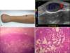
Table 1
Reported cases with eccrine spiradenoma developed on extremities
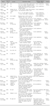
|
Reports |
Age (yr)/sex |
Location |
Characteristic finding |
Pathologic diagnosis; imaging findings |
Treatment |
|
Alfonso-Trujillo et al. (2009) |
17/F |
Leg |
0.3 to 0.5 cm sized, tender, nodular lesions of 7 years' duration. Zosteriform pattern |
Eccrine spiradenoma; - |
Surgical excision |
|
Altinyazar et al. (2003) |
32/F |
Leg |
Multiple, skin-colored, grouped, firm, tender, papulonodular lesions of 2 years' duration. Zostiform pattern |
Eccrine spiradenoma; - |
- |
|
Bedlow et al. (1999) |
19/F |
Multiple (Rt. arm, Rt. leg, Rt. side of trunk) |
15 year's duration, Tender blue-red dermal and subcutaneous nodules. Linear |
Eccrine spiradenoma; - |
- |
|
Blanchard et al. (1981) |
24/F |
Leg |
0.2 to 1 cm sized nodules for almost 12 years. Linear pattern. Asymptomatic |
Eccrine spiradenoma (Linear eccrine nevus with comedones); - |
- |
|
Bourrat et al. (1992) |
16/F |
Arm, shoulder |
Segmental lesions with the pattern of Blaschko's lines since 5 years ago. |
Eccrine spiradenoma; - |
- |
|
Brahim et al. (2009) |
75/M |
Rt. shoulder |
A well-defined, motile, firm 5.0×4.0 cm sized nodule. Tender in pressure. A 6-year history of a recurrent mass that was removed 2 years ago |
Eccrine spiradenoma, recurrent malignant eccrine spiradenoma; - |
Surgical resection |
|
Braun-Falco et al. (2003) |
23/F |
Multiple (Lt. lower leg) |
1.5 cm sized multiple nodules almost covering the frontal aspects of lower leg for almost 8 years without obvious change. Linear. Occasionally tender |
Eccrine spiradenoma (focal malignant transformation); - |
Surgical resection |
|
Criton et al. (1996) |
11/M |
Multiple (Lt. arm, Lt. leg, chest) |
0.4 to 1.0 cm sized painful tender skin colored papule of duration of 4 years. Zostiform pattern |
Eccrine spiradenoma; - |
- |
|
Englander et al. (2011) |
55/M |
Multiple (Rt. chest, Rt. arm) |
0.5 to 6.0 cm sized multiple, well-circumscribed, subcutaneous, blue-grey nodules in dermatomal distribution of 20 years' duration. Painful |
Eccrine spiradenoma; - |
Surgical resection |
|
Fahy et al. (1987) |
37/F |
Rt. Hand |
A 3.5×2.0 cm sized firm, non-tender swelling, of two years' duration, situated just distal to the right anatomical snuff-box |
Eccrine spiradenoma; - |
Surgical excision |
|
Han et al. (2007) |
24/F |
Multiple (face, neck, chest, extremities) |
0.3~2.0 cm sized well-defined nodules. Zosteriform. Painless |
Eccrine spiradenoma; MRI, low SI in T1WI, high SI in STIR |
- |
|
Hashimoto et al. (1966) |
15/F |
Forearm |
Segmental patterned lesions with 7 years' duration. Asymptomatic |
Eccrine spiradenoma; - |
- |
|
Hemalatha et al. (2015) |
80/F |
Rt. upper thigh |
A 1.75×0.75 cm sized painless raised blue to blackish lesion on thigh. Non-tender with smooth surface |
Eccrine spiradenoma; - |
Surgical excision |
|
Ikeya et al. (1987) |
37/F |
Face, trunk, arm |
0.1 to 0.8 cm sized multiple papular lesions of 30 years' duration. Linear pattern |
Eccrine spiradenoma; - |
- |
|
Jin et al. (2008) |
34/F |
Lt. upper arm |
A 1.8×1.7×1.1 cm sized firm nodular mass on the distal portion of the left upper arm, of approximately 5 years' duration. Painful |
Eccrine spiradenoma; US, well-defined lobulating mass, vascularity(+), heterogeneous hypoechogenicity |
Surgical excision |
|
Laura et al. (2011) |
55/M |
Multiple (Rt. arm, Rt. chest) |
0.5 to 6 cm sized multiple, well-circumscribed, subcutaneous, blue-grey nodules of 20 years' duration. Painful |
Eccrine spiradenoma; - |
Surgical excision |
|
Martinez et al. (1992) |
48/F |
Multiple (Lt. leg) |
0.2 to 0.6 cm sized multiple papulonodular blue-pink colored lesions appeared 18 years earlier. Asymptomatic. Tender with a firm, elastic consistency |
Multiple linear cylindromas overlapping features with eccrine spiradenoma; - |
Regular follow-up |
|
Nath et al. (2009) |
14/M |
Rt. lower leg |
A 0.5 to 1.0 cm sized multiple nodules on the posterior aspect of the entire length of the right lower limb since 2 years of age. Linear pattern (Blashkoid distribution). Painful |
Eccrine spiradenoma with chondroid syringoma; US, dermal tumors, normal vessels of the lower limb, no connection with the underlying vessels and the tumors |
Surgical excision |
|
Noto et al. (1994) |
16/F |
Multiple (Rt. leg, Rt. face, Rt. neck, Rt. trunk) |
0.5 to 5.0 cm sized nodules, observed since birth. Bluegray to bright red colored painful papulonodular lesions. Linear nevoid pattern. Painful |
Linear eccrine spiradenoma; - |
- |
|
Ohtsuka et al. (2002) |
47F |
Multiple (Rt. finger, Rt. hand, Rt. forearm) |
0.7 to 2.0 cm sized linear, nontender lesions of 30 years' duration. Localized pattern |
Eccrine spiradenoma; - |
Surgical excision |
|
Shaikh-Naidu et al. (2003) |
34/F |
Multiple (Rt. forearm) |
10×2 cm area of 12 subcutaneous nodules with overlying cutaneous scar and bluish discoloration. Previously sustained a superficial abrasion. Linear. Mild discomfort |
Eccrine spiradenoma; - |
Surgical excision |
|
Siegel et al. (2008) |
40/M |
Rt. hand |
A 6.0×4.0 cm sized slightly mobile, painless mass of 20 years' duration. No lymphadenopathy |
Giant eccrine spiradenoma; MRI, high heterogenous SI in T1WI |
Surgical resection |
|
Tsur et al. (1981) |
35/M |
Multiple (Lt. arm) |
1 to 5 cm size. 26 years of duration. Bluish and shiny with congested blood vessels. Some tumors attached by short broad stalk. Linear pattern. Painful |
Eccrine spiradenoma; - |
Surgical resection |
|
Yamakoshi et al. (2008) |
76/M |
Rt. upper arm |
A 5.0×3.4×2.6 cm sized pale red pedunculated tumor with erosive surface. No symptom except for bleeding |
Giant vascular eccrine spiradenoma; Enhanced CT, high-density nodules in the peripheral region and the central region with low density and no enhancement |
Surgical excision |
ACKNOWLEDGMENT
This study was supported by grants of the National Research Foundation of Korea (NRF), funded by the Ministry of Science, ICT & Future Planning (NRF-2017R1A2B4006252), Korea Healthcare technology R&D project, funded by Ministry of Health & Welfare, Republic of Korea (HI17C0597), and the Hallym University Research Fund (HURF-2017-35).
References
1. Park YS, Lee HE, Bang DS. A Case of Eccrine Spiradenoma. Korean J Dermatol. 1983; 21:483–487.
2. Ishikawa M, Nakanishi Y, Yamazaki N, Yamamoto A. Malignant eccrine spiradenoma: a case report and review of the literature. Dermatol Surg. 2001; 27:67–70.

3. Balaban M, Idilman IS, Unal O, Dumlu EG, Yazgan A, Ipek A. Sonographic and sonoelastographic findings of a rarely seen soft tissue tumor: eccrine spiradenoma. J Med Ultrason (2001). 2015; 42:587–590.
4. Kim MH, Cho E, Lee JD, Cho SH. Giant vascular eccrine spiradenoma. Ann Dermatol. 2011; 23:Suppl 2. S197–S200.
5. Jin W, Kim GY, Lew BL, Yang DM, Kim HC, Ryu JK, et al. Sonographic findings of an eccrine spiradenoma: case report and literature review. J Ultrasound Med. 2008; 27:813–818.


 PDF
PDF ePub
ePub Citation
 Print
Print


 XML Download