

 PDF
PDF ePub
ePub Citation
 Print
Print


Dear Editor:
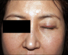
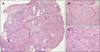
A 46-year-old woman presented with linear erythematous papules that developed from a 20-year-old scar on her left upper eyelid 2 weeks earlier. She had undergone an upper eyelid blepharoplasty 20 years earlier. She was treated with a topical steroid for 1 week, without improvement. She felt some discomfort while opening her eyes. On physical examination, erythematous, firm, non-tender papules were seen along the blepharoplasty scar on her left upper eyelid (Fig. 1). She was diagnosed with precursor B cell lymphoblastic leukemia in 2012 and achieved complete remission after allogeneic peripheral blood stem cell transplantation. For a diagnosis and exclusion of leukemia cutis, we performed a skin biopsy. Histological examination showed a dense, non-caseating granulomatous infiltration throughout the dermis, with numerous epithelioid cells. There were no atypical lymphoid cells or immature granulocytes (Fig. 2). These histological findings were consistent with sarcoidosis. A chest X-ray and laboratory studies showed no evidence of systemic sarcoidosis. After 4 months, the lesion improved spontaneously without any treatment.
Sarcoidosis is a multiorgan granulomatous disease with unknown etiology1. Scar sarcoidosis is a rare form of cutaneous sarcoidosis in which pre-existing scars are infiltrated by non-caseating granulomas. Scar sarcoidosis can develop after various events; although there are a few reports on scar sarcoidosis developing after blepharoplasty2. Moreover, there has been no report on scar sarcoidosis occurring on an old blepharoplasty scar in a leukemia patient after a surgical procedure. The patient had blepharoplasty on both upper eyelids, but the sarcoidosis appeared only on the left side. We don't know the exact mechanism for this, but different pre-operation state of upper eyelids, surgeon's skill or different post-operation care could be the reason for the asymmetric appearance of scar sarcoidosis. Similar to our case, Mantese et al.3 reported the patients who was diagnosed with scar sarcoidosis that developed only part of the scars. Although the patient had blepharoplasty, hysterectomy and caesarean, she had sarcoidosis on left upper eyelid and on the left lateral side of infra-umbical scar.
There have been several reports on sarcoidosis related to hematological malignancies. Among these, only four reports described sarcoidosis that developed after acute leukemia, after a period ranging from 11 months to 17 years4. There are several hypotheses to explain the relationship between acute leukemia and sarcoidosis. First, the granulomatous inflammation of sarcoidosis may develop in reaction to the tumor-associated antigens found in acute leukemia2. Second, the transmission of sarcoidosis or sarcoidosis-inducing pathogens via bone marrow transplantation may be associated with the development of sarcoidosis5. Scar sarcoidosis can resolve slowly and spontaneously6, although many treatments have been used for cutaneous sarcoidosis. For patients with only cutaneous sarcoidosis, with no systemic involvement, systemic treatment is not necessary and topical and intralesional steroids can be helpful. When there is systemic involvement, systemic treatment with hydroxychloroquine, prednisolone, or me thotrexate can be helpful.
This case is a rare occurrence of sarcoidosis that developed on an old upper blepharoplasty scar in a leukemia patient. We suggest a skin biopsy to exclude scar sarcoidosis when firm papules appear on a scar in leukemia patients.
 XML Download